

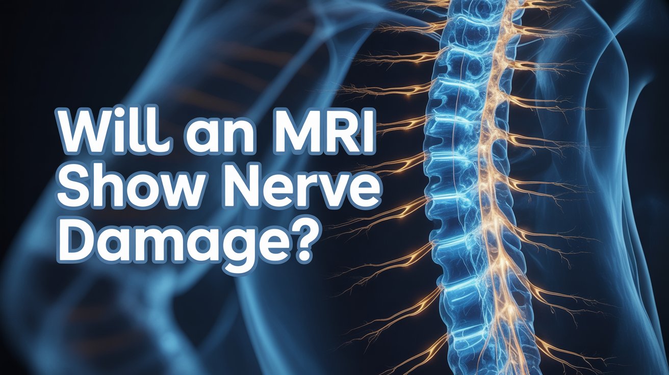
Will an MRI Show Nerve Damage?

Numbness, tingling, weakness, or burning pain are symptoms that drive thousands of people to ask whether an MRI will find the answer. The short answer is: often yes — but it depends on what type of nerve damage is involved and where it is in the body.

This guide explains what an MRI can and cannot show for nerve damage, when other tests are needed, and what symptoms should prompt you to seek assessment promptly.


Can an MRI Show Nerve Damage?

Yes. An MRI can detect nerve damage in many of the most common clinical situations — particularly where a nerve is being compressed, inflamed, or where surrounding structures are causing the problem.

Scan.com UK explains that MRI’s multiple views from different angles allow the easy detection of any displacement or protrusions putting pressure on a nerve, or anomalies in the area where the nerve exits the spine. It can also visualise the nerves themselves, along with conditions affecting them such as swelling or shrinkage.

What MRI does particularly well is show the structures surrounding nerves — the discs, vertebrae, ligaments, and soft tissues — in fine detail. When any of these press on, pinch, or damage a nerve, the MRI reveals it clearly. It also shows the spinal cord and nerve roots with a level of detail no X-ray can match.

Where MRI has limits is in measuring how well a nerve is actually working. It shows anatomy — what the nerve looks like and what is pressing on it — but not function. For that reason, it is sometimes used alongside other tests to build a complete picture.


What Types of Nerve Damage Does MRI Show Best?

MRI is most informative for nerve damage that has a clear structural or inflammatory cause. Here are the situations where it performs best.

Compressed spinal nerve roots

This is one of the most common reasons MRI is arranged for nerve symptoms. When a herniated disc, bone spur, or thickened ligament presses on a nerve root as it exits the spine, the MRI shows exactly which level is affected and how severely. This is the underlying cause of sciatica, cervical radiculopathy, and many cases of arm or leg pain, weakness, and numbness.

Spinal stenosis

Narrowing of the spinal canal compresses the spinal cord or multiple nerve roots simultaneously. MRI shows the degree of narrowing and which nerves are affected, informing decisions about treatment and surgery.

Multiple sclerosis (MS)

MS causes demyelination — damage to the protective sheath around nerve fibres. These lesions appear as bright white spots on T2-weighted MRI sequences, both in the brain and spinal cord. MRI is the primary tool for diagnosing and monitoring MS.

Spinal cord damage

Injury, inflammation, or reduced blood supply to the spinal cord itself — called myelopathy — produces characteristic changes on MRI that guide treatment urgently.

Brain-related nerve damage

Stroke, traumatic brain injury, and brain tumours can all disrupt nerve pathways. MRI of the brain identifies these structural causes with precision.


What Is MR Neurography?

For peripheral nerve damage — affecting the nerves beyond the spine — a specialist technique called MR neurography (MRN) provides significantly more detail than a standard MRI.

Research published in PMC confirms that MRN can detect small patches of inflammation in peripheral nerves and is valuable when standard MRI and nerve conduction studies have produced inconclusive results. It is particularly useful for assessing nerve lesions after trauma, planning surgery, and diagnosing entrapment neuropathies — such as carpal tunnel syndrome, ulnar nerve entrapment at the elbow, and sciatic nerve compression.

MRN uses high-resolution T2-weighted sequences with fat suppression to isolate the nerve signal from surrounding tissue, making individual peripheral nerves visible in a way that standard MRI cannot achieve. It is not available at every NHS centre and is more commonly accessed privately or through specialist neurological referral.


What Can MRI Not Show for Nerve Damage?

MRI shows structure, not function. This is its most important limitation when investigating nerve damage.

Howe Law’s clinical summary on MRI and nerve damage explains that MRI primarily shows the morphology of nerves — what they look like physically — but cannot provide definitive information about how well the nerve is working. A nerve can appear structurally normal on MRI while still conducting signals poorly or causing significant symptoms.

Specific types of nerve damage that standard MRI often cannot detect include:

Diabetic peripheral neuropathy

This condition damages small nerve fibres throughout the body, causing pain, numbness, and tingling — but often without the structural changes that MRI is designed to pick up. Howe Law notes that neuropathy secondary to diabetes may not always be detectable through standard MRI techniques.

Small fibre neuropathy

This affects the smallest sensory nerve fibres and typically requires a skin biopsy for diagnosis — MRI does not reliably show these microscopic changes.

Early-stage nerve damage

In the very early stages of compression or injury, structural changes may be too subtle to appear on MRI even while symptoms are already present.

A normal MRI result does not rule out nerve damage. If your symptoms are consistent with nerve involvement but your MRI is unremarkable, further investigation using functional tests is the appropriate next step.


What Other Tests Are Used to Diagnose Nerve Damage?

When MRI alone is not sufficient — or when the clinical question is about how well a nerve is working rather than what is pressing on it — two additional tests are commonly used.

Nerve Conduction Study (NCS)

A nerve conduction study measures the speed and strength of electrical signals travelling through peripheral nerves. North Bristol NHS Trust explains that it assesses the function of the sensory and motor nerve pathways, and is used when a patient is experiencing numbness, weakness, or changes in sensation to the hands or feet.

During the test, small electrical pulses are applied to the surface of the skin over a nerve. The response is recorded at another point along the nerve’s path. The speed and strength of that response reveal whether the nerve is conducting signals normally or is damaged.

Electromyography (EMG)

EMG measures the electrical activity inside muscles, revealing whether abnormal patterns are the result of nerve damage or a muscle disorder. King’s College Hospital NHS confirms that NCS and EMG together are useful for diagnosing entrapment syndromes such as carpal tunnel syndrome, polyneuropathy (for example in diabetic patients), and other peripheral nervous system disorders.

A fine needle electrode is inserted into the muscle being examined. The electrical activity it records — both at rest and during contraction — tells the clinician whether the nerve supplying that muscle is functioning normally.

NCS and EMG complement MRI rather than replace it. MRI shows the structural cause; NCS and EMG show the functional impact. Used together, they give clinicians the clearest possible picture of what is happening and why.


When Should You Get an MRI for Nerve Damage Symptoms?

If you are experiencing numbness, tingling, weakness, or pain that radiates from your neck or back into an arm or leg, an MRI is usually the right first-line imaging investigation. These symptoms suggest a nerve root is being compressed somewhere along the spine, and MRI is the most effective way to find out where and why.

Symptoms that warrant prompt assessment include pain, numbness, or tingling that travels down one arm or leg, unexplained muscle weakness, a sensation of burning or electric shocks in the limbs, and loss of coordination or balance.

Certain symptoms should be treated as urgent. Sudden or rapidly worsening weakness in both legs, loss of bladder or bowel control, or numbness in the groin and inner thighs (saddle anaesthesia) are red flag signs that require immediate emergency attention — call 999 or go to A&E.

For symptoms that are less acute — persistent but not rapidly worsening — a same-day private GP assessment allows for prompt clinical evaluation, appropriate investigation, and referral if needed. Our private GP consultation is available with no waiting list, and our full health check-up can help identify contributing factors such as diabetes or nutritional deficiencies that may be causing or worsening nerve symptoms.


Frequently Asked Questions

  • Can an MRI detect a pinched nerve?

Yes. MRI is one of the best tests for identifying a pinched nerve — it clearly shows if a disc, bone spur, or other structure is compressing a nerve root, and at which level of the spine.

  • Will an MRI show sciatica?

MRI can show the cause of sciatica — most commonly a herniated disc pressing on the sciatic nerve root. It does not diagnose sciatica itself, but reveals the structural problem driving the symptoms.

  • Can an MRI show nerve damage in the neck?

Yes. A cervical spine MRI shows the nerve roots exiting the neck, any disc herniation or stenosis compressing them, and the spinal cord itself. It is the standard investigation for neck-related arm pain, numbness, and weakness.

  • What is the best test to diagnose nerve damage?

It depends on the type. MRI is best for structural causes such as disc compression and MS. Nerve conduction studies and EMG are best for assessing peripheral nerve function. In many cases, a combination of tests gives the clearest diagnosis.

  • Can an MRI miss nerve damage?

Yes. MRI shows structure, not function, so it can appear normal even when nerve damage is present — particularly in diabetic neuropathy, small fibre neuropathy, and early-stage compression. A normal MRI does not rule out nerve damage if symptoms persist.

How Long Does a Full Spine MRI Take?

A full spine MRI covers a lot of ground — from the base of your skull all the way down to your lower back. That means it takes considerably longer than a scan of just one section. If you know what to expect time-wise before you arrive, the appointment is far less daunting.

This guide covers how long the scan takes, how it compares to individual section scans, what happens during the appointment, and what a full spine MRI can show.


How Long Does a Full Spine MRI Take?

A full spine MRI typically takes between 60 and 90 minutes inside the scanner. You should allow up to two hours for your total time at the clinic, including preparation, changing, safety checks, and any post-scan wait if contrast dye is used.

Healthline confirms that a cervical spine scan takes around 20 to 45 minutes, and a lumbar spine scan takes around 30 to 60 minutes. A full spine scan — covering all three sections — requires each region to be imaged separately with its own sequences, which is why the total time is significantly longer than any individual section alone.

Harrogate and District NHS Foundation Trust notes that while most MRI scans take around 30 minutes, some take up to 90 minutes — and a full spine scan sits firmly at the longer end of that range.


How Does This Compare to a Single Section Scan?

Here is a quick breakdown of typical scan times by spinal region:

  • Cervical spine (neck): 20 to 45 minutes
  • Thoracic spine (mid-back): 30 to 45 minutes
  • Lumbar spine (lower back): 30 to 60 minutes
  • Full spine (all three sections): 60 to 90 minutes

Each section requires the coil to be repositioned and a new set of sequences to be run, which is why three sections together do not simply equal three times as long — but they do add up considerably.


What Happens During a Full Spine MRI?

You lie flat on a padded bed and enter the scanner head-first. University Hospital Southampton NHS confirms that you will be positioned in the centre of the scanning bed and will need to lie very still throughout to achieve the best quality imaging.

A coil — a flat receiver pad — is placed over the section of your spine being imaged. For a full spine scan, the radiographer will reposition this coil between sections, which adds a small amount of time between imaging runs. You will be moved out briefly, the coil adjusted, and then moved back in for the next section.

The scanner produces loud tapping and knocking sounds throughout. University Hospital Southampton advises that you may also feel your body temperature increase slightly and experience tingling in your hands and feet — both of which are normal sensations caused by the magnetic field. You will be given an emergency buzzer to hold at all times, and the radiographer can hear you via a built-in microphone system between sequences.

For a lumbar spine scan specifically, Ezra’s clinical guide notes that patients are often positioned feet-first rather than head-first, which can make the experience more comfortable for those who feel anxious about enclosed spaces — as the head and upper body remain outside the tunnel for that section.


What Can Make a Full Spine MRI Take Longer?

Several factors can extend the time beyond the typical 60 to 90 minutes.

Contrast dye

If your clinician has requested contrast-enhanced imaging, a gadolinium-based dye is injected through a cannula in your arm. A second set of images is taken after injection, adding around 15 to 30 minutes to the scan time.

Movement

The MRI machine is sensitive to any movement. UK Spine Centre confirms that it is crucial for patients to remain still throughout the scanning process to ensure clear images. If movement blurs a sequence, it must be repeated — which extends the total time.

Number of sequences

The number of image sequences ordered by your clinician varies depending on what they are investigating. More detailed or complex clinical questions require more sequences, which adds time.

Patient size

Larger body habitus can require adjustments to coil positioning and signal strength settings, which occasionally adds time to individual sequences.


What Does a Full Spine MRI Show?

A full spine MRI gives clinicians a comprehensive view of the entire spinal column — from the vertebrae and discs to the spinal cord, nerve roots, and surrounding soft tissue.

UK Spine Centre outlines the range of conditions a spinal MRI can identify, including:

Disc herniation and sciatica

A herniated disc pressing on a nerve root is one of the most common reasons for a spinal MRI. The scan shows exactly which disc is affected and how severely it is compressing the nerve.

Spinal stenosis

Narrowing of the spinal canal — whether from disc degeneration, bone spurs, or thickened ligaments — compresses the spinal cord or nerve roots and shows up clearly on MRI.

Nerve compression

Wherever a nerve is being pinched or squeezed along the length of the spine, MRI identifies the cause and the level of involvement.

Spinal cord abnormalities

Conditions affecting the cord itself — including MS lesions, inflammation, or areas of poor blood supply — are visible on MRI in a way no other imaging method can match.

Tumours

Both primary spinal tumours and metastatic disease (cancer that has spread to the spine) can be identified and characterised using MRI.

Fractures and infections

Vertebral fractures — including stress or compression fractures that may not appear on X-ray — and bone infections (osteomyelitis) show up on MRI with detail that guides treatment decisions.

Post-surgical assessment

If you have had previous spinal surgery, MRI can assess whether there is scarring, recurrent disc herniation, or infection around the operated area.


How to Prepare for a Full Spine MRI

Preparation for a full spine MRI is straightforward. Ezra’s guidance confirms that unless told otherwise, you can eat, drink, and take your usual medications as normal on the day. No fasting is required for a standard spine MRI without contrast. If contrast dye has been requested, your appointment letter will specify any fasting instructions.

Remove all jewellery, piercings, and metal accessories before attending. Wear loose, comfortable clothing without metal zips, buttons, or fasteners — a t-shirt and tracksuit bottoms work well. You may be asked to change into a hospital gown on arrival.

Tell the MRI team in advance about any metal implants, surgical clips, or devices in your body, including spinal hardware such as rods, screws, or cages from previous surgery. Most modern spinal implants are MRI-compatible, but this must be confirmed beforehand.

If you are anxious about lying still for 60 to 90 minutes or feel uncomfortable in enclosed spaces, speak to your GP before the appointment. A mild sedative can be prescribed to help you remain calm, but it must be arranged in advance as it cannot be prescribed on the day.

If you are experiencing back, neck, or leg symptoms and are unsure whether a spine MRI is the right investigation, our full health check-up gives you a thorough clinical assessment to help determine the most appropriate next step.


Frequently Asked Questions

  • Is a full spine MRI the same as three separate scans?

Effectively yes — each section (cervical, thoracic, lumbar) is imaged with its own sequences. It is done in one appointment, but the coil is repositioned between sections and each region is scanned individually.

  • Can I move between sections of a full spine MRI?

You will be briefly moved out of the scanner between sections for coil repositioning. During each imaging run, you must remain completely still to avoid blurred images requiring repetition.

  • Will my whole body go inside the scanner for a full spine MRI?

For the cervical and thoracic sections, most of your body enters the tunnel. For the lumbar section, entry is often feet-first, meaning your head and upper body can remain outside.

  • How long after a full spine MRI will I get results?

A radiologist analyses the images and sends a report to your referring doctor, typically within a few working days. Your doctor will then contact you to discuss the findings.

  • Do I need contrast dye for a full spine MRI?

Not always. Contrast is used when the clinical team needs clearer detail — for example, when investigating tumours, infection, or inflammation. Your appointment letter will confirm whether it has been requested.

What to Wear for an MRI Scan

Choosing the right outfit for an MRI takes about two minutes of planning and can make your appointment considerably more comfortable. The main rule is simple: avoid metal. Here is everything you need to know before you arrive.


What Should You Wear to an MRI Scan?

Wear loose, comfortable clothing with no metal fastenings. This is the single most important clothing rule for an MRI, and it applies regardless of which part of your body is being scanned.

NHS Inform confirms that if you do not need to wear a gown, you should wear clothes without metal zips, fasteners, buttons, underwire bras, belts, or buckles. Surrey and Sussex Healthcare NHS Trust advises choosing thin cotton-based clothing with minimal layers, as this also helps with cooling during the scan — the magnetic field can cause a mild warming sensation in some patients.

Good choices include:

  • A plain t-shirt or cotton top with no metal press studs or zips
  • Loose tracksuit bottoms or joggers with an elasticated waistband
  • Soft, wireless sports bra or a comfortable bralette
  • Slip-on shoes or trainers without metal eyelets


What Clothing to Avoid

Certain everyday items cause problems in the scanning room and should be left at home or be prepared to be removed:

Jeans

Most jeans have metal rivets, metal zips, and metal buttons. They are one of the most common items patients are asked to remove before a scan.

Underwired bras

The metal underwire must be removed before scanning. A wireless bra or soft sports bra is a much easier choice and saves the need to change.

Belts

Any belt with a metal buckle needs to come off. Elasticated waistbands avoid this entirely.

Hoodies and jackets

Many have metal zips, metal drawstring tips, or metal poppers. A plain sweatshirt or cardigan without fastenings is a better option.

Compression garments

Some compression leggings and athletic wear contain metallic fibres in the fabric. If in doubt, opt for plain cotton instead.

Leeds Teaching Hospitals NHS Trust advises that if there is any metal on your clothing near the area being scanned, you may be asked to remove that item of clothing and a gown will be provided.


Does It Matter Which Body Part Is Being Scanned?

Yes, slightly. The area being scanned determines how much of your clothing is relevant.

For head, brain, or neck scans, your clothing from the shoulders down is generally not a concern. Focus on removing all metal from the head and neck area — hairpins, earrings, necklaces, and piercings.

For chest or cardiac scans, avoid underwired bras and anything with metal across the chest. ECG stickers are attached to the chest during cardiac MRI, so easy access matters. If you are having an ECG heart health check-up as part of your cardiac assessment, the same principle applies.

For spine, abdomen, or pelvic scans, the focus shifts to your lower half. Avoid jeans, belts, and anything with metal fastenings around the waist or hips. Loose trousers or joggers are the simplest choice.

For knee, ankle, or foot scans, most of your clothing is irrelevant to the scan itself. Wear whatever is comfortable and easy to remove from the lower leg if needed.


Will You Need to Change Into a Hospital Gown?

Sometimes — but not always. Whether you need a gown depends on which part of your body is being scanned and whether your clothing is already metal-free.

NHS Inform confirms that depending on which part of your body is being scanned, you may need to wear a hospital gown during the procedure. The Royal Marsden NHS Trust advises that when you arrive for your appointment, it is advisable to wear clothing without metal fastenings — alternatively, they can provide a hospital gown or pyjamas.

If you arrive in entirely metal-free clothing, many clinics will allow you to stay in your own clothes throughout. This is one of the reasons arriving dressed appropriately saves time and avoids any awkwardness on the day.

When a gown is provided, you will be directed to a changing room with a secure locker for your belongings. Your dignity and privacy are maintained throughout — only the area being scanned is exposed, and you remain covered otherwise.


What Jewellery and Accessories Should You Remove?

All of it. Every piece of metal jewellery and every metal accessory must be removed before you enter the scanning room, without exception.

This includes:

  • Rings, earrings, necklaces, and bracelets
  • Body piercings of any kind
  • Watches and fitness trackers
  • Hair clips, kirby grips, and metallic hairbands
  • Hearing aids
  • Glasses and spectacle frames
  • Removable dental work such as retainers or partial dentures

Royal Devon University Healthcare NHS Foundation Trust advises attending with clothing that has no clips, zips, or belt fastenings, and removing all metal accessories before the scan.

Leave valuables at home where possible. Most clinics provide secure lockers for items you do bring, but a lost or damaged item of jewellery or a watch with a damaged magnetic strip is easily avoided by not bringing it in the first place. Credit and debit cards should also be left outside the scanning room, as the magnetic field can erase or damage the card’s strip.


What About Make-Up and Cosmetics?

Make-up is a less obvious consideration, but it is worth knowing about before your appointment.

Leeds Teaching Hospitals NHS Trust specifically advises not wearing cosmetics with magnetic substances — including magnet-attached eyelashes — and ensuring any previously applied make-up of this type is fully removed on the day. Royal Devon NHS advises avoiding heavy eye make-up, as this can affect the quality of some scans.

Some mascaras, eyeshadows, and eyeliners contain iron oxide or other metallic pigments. These particles can interact with the magnetic field, causing a mild warming sensation around the eye area and occasionally affecting image quality near the head.

Magnetic eyelashes — which use tiny magnets as their adhesive mechanism — must be removed before scanning without exception. Standard press-on lashes attached with glue are fine.

Skin lotions, moisturisers, and sun cream do not pose a problem for most MRI scans. However, for cardiac MRI where ECG electrodes are attached to the chest, any lotion applied to the chest area should be washed off beforehand, as it can reduce electrode adhesion and affect monitoring quality.


Frequently Asked Questions

  • Can I wear a sports bra for an MRI?

Yes, provided it is wireless and has no metal clasps, hooks, or underwire. A soft, wireless sports bra or bralette is the ideal choice for any scan involving the chest or upper body.

  • Can I wear earrings to an MRI?

No. All earrings — including small studs — must be removed before entering the scanning room. If you have a new piercing that cannot be removed, let the MRI team know in advance so they can advise.

  • What happens if I forget and wear metal to my MRI?

You will be asked to remove the item before the scan. If it is an item of clothing that cannot easily be removed, a gown will be provided. The scan will still go ahead in most cases — it just adds a few minutes to your preparation time.

  • Can I wear my own clothes or do I have to wear a gown?

If your clothing is entirely metal-free, many clinics will allow you to stay in your own clothes. If there is any metal near the area being scanned, a gown will be provided. Arriving in suitable clothing is the easiest way to avoid needing to change.

  • Can I wear nail varnish to an MRI?

Standard nail varnish is fine for most MRI scans. Some metallic or glitter nail polishes contain metal particles and may cause a slight warming sensation, but this is uncommon. If you are having a scan of the hands or fingers, it is worth removing nail varnish beforehand to avoid any interference with image quality.

How Long Does Minor Surgery Take?

If you have been booked in for minor surgery and are wondering whether to take the whole day off work, the good news is that you probably do not need to. Most minor surgical procedures at a GP clinic are significantly quicker than people expect.

That said, the time varies considerably depending on what is being done, where on the body it is, and how straightforward the procedure turns out to be. Knowing what to expect — and how long to set aside — removes one of the most common sources of anxiety before the appointment.

If you are considering minor surgery and want to discuss your options first, our private GP consultation can help you understand what is involved before you commit to anything. Same-day appointments are available, and no referral is needed.


What Is Minor Surgery?

Minor surgery covers a range of small procedures performed under local anaesthetic in a clinic setting — without the need for a general anaesthetic, an operating theatre, or an overnight hospital stay. You are awake throughout, the area being treated is numbed, and you go home the same day.

The Private GP’s minor surgery service describes it as small-scale procedures typically used for non-life-threatening conditions such as skin lesions, small lumps, or ingrown nails — all performed under local anaesthesia in a clean clinical environment.

Common minor surgical procedures include:

Skin tag removal, mole excision, cyst removal (sebaceous and epidermoid), lipoma removal, wart and verruca treatment, abscess drainage, ingrown toenail surgery, wound suturing and closure, and removal of foreign bodies from the skin.

Most people have minor surgery either because something is causing discomfort or pain, because a skin lesion is changing and needs to be checked, or simply because they want something removed for practical or cosmetic reasons. A pre-operative consultation is always arranged before any procedure, so you have the chance to ask questions and confirm you are suitable for treatment.


How Long Does Minor Surgery Take?

Most minor surgical procedures take between 15 and 45 minutes from the moment you enter the treatment room to the moment you leave. The procedure itself is often just a fraction of that time — preparation, injecting the local anaesthetic, and post-procedure wound care account for much of the appointment.

Lancaster Medical Practice’s patient information confirms that depending on the complexity of the surgical procedure and the site of the lesion, surgery can take from around 15 minutes to 45 minutes. Tisbury Surgery’s NHS patient leaflet puts the range at 30 to 60 minutes for more involved procedures.

At The Private GP in Birmingham, our minor surgery procedures are performed under local anaesthesia and typically take 20 to 30 minutes. Most patients are in and out of the clinic well within an hour, including the time before and after the procedure itself.

The main factors that affect how long your appointment will take are the type and size of the lesion, its location on the body, the closure technique used, and how quickly the local anaesthetic takes effect. Complex lesions, those in difficult anatomical locations, and procedures requiring deeper excision naturally take longer than straightforward surface removals.


How Long Does Each Type of Minor Surgery Take?

Different procedures have different time profiles. Here is a practical breakdown.

Skin tag removal: 15 to 20 minutes. Skin tags are among the quickest procedures. They are snipped or treated at the surface, require minimal or no stitching, and heal quickly. The appointment is short and straightforward.

Cyst excision: 20 to 30 minutes. Removing a sebaceous or epidermoid cyst involves opening the skin, excising the cyst wall (to prevent regrowth), and closing the wound with stitches. The procedure itself takes 10 to 15 minutes; the rest of the time is preparation and dressing.

Mole or skin lesion excision: 20 to 45 minutes. The time depends on the size of the lesion and how wide a margin of surrounding skin needs to be removed. Smaller lesions at the surface are quicker; deeper or larger excisions take longer and may require more stitches.

Lipoma removal: 20 to 45 minutes. A lipoma is a benign fatty lump under the skin. Removing it involves an incision, careful dissection to free the lump, and closure. Larger lipomas or those in deeper tissue take longer.

Ingrown toenail surgery: 30 to 60 minutes including preparation. North Tees NHS Foundation Trust confirms that the operation itself usually takes around 10 to 15 minutes, but the total appointment time — including anaesthetic preparation, a wait for the toe to numb fully, the procedure, and dressing — brings it closer to an hour. A tourniquet is used during the procedure to minimise bleeding, and you will need someone to take you home afterwards.

Abscess drainage: 15 to 30 minutes. Draining an abscess is generally quick. The area is numbed, a small incision is made, the abscess is drained, and the wound is packed or dressed. Larger or more complex abscesses take longer.

Wound suturing: 15 to 30 minutes. Closing a wound with stitches depends on the length and depth of the wound. Simple lacerations are quick; deeper or irregular wounds requiring layered closure take more time.


What Happens During the Appointment?

Understanding the sequence of events helps make the appointment feel much less daunting. Here is what to expect from start to finish.

Before the procedure

Aylesford Medical Centre’s patient guidance explains that before surgery, your clinician will explain exactly what procedure is being done and why. You will have the opportunity to ask questions about the procedure and any alternative treatments, including what would happen if you decided not to have surgery. Provided you are happy, you will be asked to sign a consent form.

Preparation

You will be asked to expose only the area being treated — you do not need to undress completely. The skin is cleaned with an antiseptic solution to reduce infection risk.

Local anaesthetic

A very fine needle is used to inject local anaesthetic into and around the area. This causes a mild stinging sensation for a few seconds. After that, the area becomes numb and you will feel no pain during the procedure itself — only possibly some pressure or movement. The anaesthetic takes effect within a few minutes.

The procedure

The clinician performs the excision, drainage, or treatment. This is often the shortest part of the appointment. You remain awake and can talk throughout. If anything feels uncomfortable, let the clinician know immediately.

Wound closure and dressing

Depending on the procedure, the wound may be closed with stitches (dissolvable or non-dissolvable), wound glue, steri-strips, or left to heal naturally under a dressing. You will be given a dressing before you leave.

Aftercare instructions

Before you go home, your clinician will walk you through exactly how to care for your wound, when to keep it dry, when stitches need to be removed, and what signs of infection to watch for. At The Private GP, written aftercare guidance is also provided.

The local anaesthetic typically wears off around three hours after the procedure. Aylesford Medical Centre advises starting over-the-counter pain relief such as paracetamol about two hours after the procedure, before the anaesthetic fully wears off, so that it is already working when any discomfort begins.


How Long Is Recovery After Minor Surgery?

Minor surgery is specifically designed to have minimal downtime. For most people, recovery is straightforward and does not significantly disrupt daily life.

Cambridge University Hospitals’ post-operative guidance advises that after simpler procedures, patients should rest for a few hours, and after larger procedures, for a few days. You can go home the same day in almost all cases.

Returning to work. Most people with desk-based jobs can return to work the next day. If your work involves physical activity, heavy lifting, or the treated area is somewhere that clothing rubs against, you may need a few more days. Your clinician will advise based on your specific procedure and job.

Wound care. University Hospitals Sussex’s post-operative wound care guidance is clear: keep the dressing dry for at least the first 48 hours. After that, you can shower gently, letting water run over the area without soaking or rubbing it. Avoid baths and swimming until the wound is fully healed and stitches have been removed.

Stitches. Hull University Teaching Hospitals NHS Trust confirms that stitches or dressings are typically removed 7 to 14 days after the procedure. Non-dissolvable stitches need to be taken out by a practice nurse at your GP surgery — book this appointment as soon as possible after your procedure. Dissolvable stitches break down on their own, usually within 7 to 14 days, though some types may take a few weeks.

Exercise. University Hospitals Sussex advises avoiding vigorous exercise such as swimming, running, contact sports, or the gym until the stitches have been removed and the area has healed. Light walking is generally fine from the same day.

Biopsy results. If your tissue sample is sent for histological analysis — which is routine for any mole or lesion that is cut out, not just suspicious ones — results typically take 4 to 8 weeks. You will be contacted with the outcome once available.

For patients who find it difficult to travel in the days after their procedure, our home visit service is available to provide follow-up care in the comfort of your own home.


What Speeds Up or Slows Down Recovery?

Recovery time is not the same for everyone. Several factors influence how quickly your wound heals.

Location on the body. Wounds on the face and scalp tend to heal faster because of the excellent blood supply to these areas. Wounds on the lower legs, particularly in older patients, heal more slowly. Post-operative guidance from Leeds Teaching Hospitals confirms this and recommends avoiding strenuous exercise that might put strain on stitches for the first two to three weeks.

Smoking. Smoking significantly delays wound healing, increases the risk of infection, and can contribute to a more prominent scar. Leeds Teaching Hospitals specifically advises reducing or stopping smoking during recovery. If you would like support with this, speak to your practice nurse or call the NHS Smoking Helpline.

Diabetes. Diabetes can impair wound healing and increase infection risk. If you have diabetes, tell your clinician before your procedure so they can give you tailored aftercare advice and monitor your healing more closely. Our full health check-up can help you understand your overall health picture including any factors that might affect surgical recovery.

Blood thinners. Medications such as aspirin, warfarin, and clopidogrel can increase bleeding during and after the procedure. Do not stop taking these without specific advice from your clinician — this will be discussed at your pre-operative consultation.

Nutrition. Leeds Teaching Hospitals’ guidance notes that good nutrition is always important but becomes even more so when the body is recovering from surgery. Adequate protein intake in particular — from fish, chicken, dairy products — provides the building blocks the body needs for tissue repair.

Signs of infection to watch for. Some redness and swelling around the wound in the first few days is entirely normal. Contact your GP promptly if the wound becomes increasingly red, hot, swollen, or painful after the third day, or if you notice yellow or green discharge, an unpleasant smell, or feel generally unwell with a temperature. Infection is uncommon when proper aftercare is followed, but catching it early makes treatment straightforward.


Frequently Asked Questions

  • Do I need a consultation before minor surgery at The Private GP?

Yes. A pre-operative consultation is required before any minor surgical procedure. This allows your clinician to assess the lesion or condition, confirm your suitability for the procedure, review your medical history and any medications, and discuss the options and risks with you. You can book your pre-operative consultation directly — no GP referral is needed.

  • Will I need time off work after minor surgery?

For most desk-based jobs, you will not need more than a day off — and some patients return to work the same afternoon. If your job involves physical activity, heavy lifting, or the treated area is at risk of being knocked or rubbed, your clinician will advise you on the appropriate timeframe. Most people are back to their usual routine within 24 to 48 hours.

  • Can I drive myself home after minor surgery?

For most procedures, you can drive yourself home. However, if the procedure involves your hand, foot, or toe — or if the local anaesthetic affects your ability to use the relevant limb safely — you should arrange for someone else to take you home. North Tees NHS advises that after ingrown toenail surgery in particular, you should not drive on the day of the procedure. Your clinician will confirm this at your appointment.

  • When will my stitches be removed?

Non-dissolvable stitches are typically removed 7 to 14 days after your procedure, depending on where on the body they are. Stitches on the face may be removed earlier — around 5 to 7 days — while those on the body or legs may stay in for up to 14 days. Your clinician will tell you the exact timeframe on the day of your surgery. Book the appointment with your practice nurse as soon as possible after your procedure.

  • How long until I get biopsy results after minor surgery?

If your tissue sample is sent for histological analysis, results typically take 4 to 8 weeks. This is routine for all lesions that are excised and does not necessarily mean there is any cause for concern. You will be contacted with the results once they are available, and a follow-up appointment will be arranged if needed to discuss them.

Can You Eat Before an MRI Scan?

You have an MRI booked and you are wondering whether you need to skip breakfast. For most people, the answer is no. But the rules vary depending on what is being scanned and whether contrast dye is involved — and getting this wrong could mean your scan has to be postponed and rescheduled.

This guide gives you a clear answer based on your scan type, explains why fasting is sometimes necessary, and covers everything else you need to know to walk into your appointment prepared.


Can You Eat Before an MRI? The General Rule

For most standard MRI scans, you can eat, drink, and take your usual medication as normal beforehand. No special preparation is needed.

The NHS confirms that the hospital where you are having your MRI will tell you if there is anything you need to do before the scan. If your appointment letter does not mention fasting or dietary restrictions, you do not need to change anything.

Cambridge University Hospitals’ patient information is explicit: on the day of your MRI scan, you should be able to eat, drink, and take any medication as usual, unless you have been advised otherwise. Leeds Teaching Hospitals confirms the same — in most cases you do not need to do anything to prepare for the scan and can eat and drink as normal.

This means that if you are having a brain MRI, a spinal MRI, a knee scan, a shoulder scan, or any other joint or extremity scan, you can have your breakfast, lunch, or dinner as normal before you arrive. There is no need to rush out of the house on an empty stomach.

The one golden rule that applies to every MRI appointment: read your appointment letter carefully and follow any specific instructions it contains. If the letter says to fast, fast. If it says nothing about food, eat as normal. If you are ever unsure, ring the radiology department before the day.


When Do You Need to Fast Before an MRI?

Fasting is required for a specific set of MRI scans — those where food in the digestive system would blur the images, or where contrast dye or sedation creates a nausea or safety risk.

Ezra’s clinical guide on MRI preparation explains that although many MRI scans can proceed without prior fasting, a sub-selection of scans require it. Here are the main situations where you will be asked to fast.

Abdominal and pelvic MRI

When the scanner is imaging your abdomen — including the liver, pancreas, kidneys, gallbladder, and surrounding organs — food in your digestive system causes a problem. Eating triggers peristalsis, the involuntary muscle contractions that move food through your gut. These movements create motion on the images, known as artefacts, which blur the scan and can obscure abnormalities. Fasting slows this activity down, giving the radiologist a much clearer picture. University Hospitals Plymouth’s liver MRI patient information confirms that patients must not eat anything for 4 hours before their appointment, though they can drink water or squash in that time.

Liver, pancreas, and gallbladder scans

As well as the motion artefact problem, eating causes your gallbladder to contract and shrink, making it harder for the radiologist to assess it properly. Fasting keeps the gallbladder distended and clearly visible.

MRCP (Magnetic Resonance Cholangiopancreatography)

This is a specialist MRI used to image the bile ducts, gallbladder, and pancreatic duct system. The fasting requirement for MRCP is particularly strict, often 6 to 8 hours, because the scan requires the digestive system to be as empty as possible to avoid fluid secretions interfering with the images.

MRI with contrast dye

When a gadolinium-based contrast agent is injected during the scan, it can occasionally cause nausea as a side effect. GetScanned’s fasting guide explains that fasting for 4 to 6 hours before a contrast scan is usually required to reduce the risk of vomiting while you are lying flat inside the scanner — a potentially serious situation as stomach contents could enter the airway. Your radiology department will specify the exact fasting period in your appointment instructions.

Cardiac MRI

For certain cardiac MRI examinations, food restriction and caffeine avoidance are both required. Craft Body Scan’s clinical preparation guide confirms that caffeine — including coffee (regular and decaf), tea, energy drinks, and chocolate — must be avoided for 24 hours before a cardiac MRI. Caffeine increases heart rate, making it harder to capture clear, stable images of the heart. If you are investigating cardiac symptoms, our ECG heart health check-up is a useful first step before moving to advanced cardiac imaging.

Scans requiring sedation or general anaesthetic

If you are having a sedative to manage claustrophobia or anxiety, or if your scan requires a general anaesthetic, fasting is essential for safety. When sedated, the protective reflexes that prevent aspiration — inhaling stomach contents if you vomit — are reduced. Fasting ensures the stomach is empty if this occurs. The Royal Marsden NHS Trust confirms that all instructions will be included in your appointment letter, and it is important to read it carefully.


How Long Do You Need to Fast Before an MRI?

When fasting is required, the standard period is 4 hours — no food, though water is usually still permitted.

NHS Highland’s patient information for fasting MRI scans gives a clear instruction: do not eat any food or drink any fluids for 4 hours before your appointment time. Cambridge University Hospitals confirms that in some cases you may be asked not to eat or drink anything for up to four hours before the scan.

For MRCP and some gastrointestinal MRI scans, the fasting period may be longer — up to 6 to 8 hours — so that the digestive system is as empty and still as possible. Your appointment letter will specify the exact time.

If you have been asked to fast and are finding it difficult, try to book an early morning appointment where possible. Sleeping through most of the fasting window makes it considerably more manageable. If you have diabetes or another condition that makes fasting medically complicated, contact the radiology department before your appointment — this is covered in more detail below.


Can You Drink Water Before an MRI?

In most cases, yes — water is permitted before an MRI, even during a fasting period.

GetScanned’s fasting preparation guide confirms that water is often allowed before an MRI and that staying hydrated is actually recommended, as it can make vein access easier if a contrast injection is needed. University Hospitals Plymouth’s liver MRI leaflet specifically tells patients they may drink water or squash in the 4 hours before their scan, even though food is not permitted.

Some pelvic MRI scans actually require you to arrive with a full bladder. In those cases, you will be asked to drink a specified amount of water before your appointment and not to urinate until after the scan is complete. Your appointment letter will tell you if this applies to you.

One important exception: caffeine. For cardiac MRI specifically, Craft Body Scan’s guide confirms that coffee (including decaffeinated), tea, energy drinks, cola, and chocolate must all be avoided for 24 hours before the scan. These contain caffeine which raises the heart rate and makes clear cardiac imaging significantly harder. If in doubt, stick to water for the day before a cardiac MRI.

A practical note worth keeping in mind: MRI scans can last between 30 minutes and 90 minutes, and you cannot leave the scanner partway through. It is sensible to limit how much you drink in the hour or two immediately before your appointment, simply for your own comfort.

After your scan, GetScanned advises drinking plenty of water — especially if you had contrast dye — to help your kidneys flush the gadolinium from your system efficiently.


Can You Take Medication Before an MRI?

For most patients, the answer is yes — continue taking all your regular medication as normal before an MRI.

Cambridge University Hospitals and Leeds Teaching Hospitals both confirm that in most cases you should eat, drink, and take your medication as usual. Royal United Hospital Bath’s MRI patient leaflet states that you should continue to take all your normal medication, with one important exception.

Diabetic patients need to take extra care, particularly those taking insulin or metformin. If you have diabetes and your MRI requires fasting, the interaction between fasting and your diabetes medication needs to be managed carefully. NHS Highland’s fasting MRI guidance includes specific diabetic instructions: if you take diabetes medication with food as prescribed, adjustments will be needed around the fasting window. Contact the radiology department before your appointment to discuss this — most NHS trusts aim to book diabetic patients for early-morning slots to minimise the fasting period.

If you have kidney disease and are taking metformin, this is particularly important to mention before any MRI involving contrast dye. GetScanned’s guide advises that metformin can pose a risk of complications in individuals with kidney problems when contrast is used. Our private blood tests can check your kidney function promptly if this is a concern before your scan.

Sedatives for claustrophobia must be prescribed by your GP before your appointment — the radiology department cannot prescribe medication on the day. If you think you might struggle with the enclosed space, speak to your GP well in advance. If you do take a sedative, you will need someone to drive you home afterwards, as you will not be able to drive for 24 hours.


Other Things to Know Before Your MRI Appointment

Beyond the question of food, there are a few other practical points worth knowing before your scan.

Metal

Remove all jewellery, piercings, watches, and accessories before you arrive. Avoid underwire bras, belts with metal buckles, and clothing with metal zips or buttons near the area being scanned. Leeds Teaching Hospitals suggests arriving in a t-shirt, jogging bottoms, and a sports bra if possible, to avoid having to change into a hospital gown.

Make-up

Some mascaras and metallic make-up products contain iron particles that can cause interference with image quality or feel warm during the scan. NHS Highland’s preparation guidance specifically advises patients not to wear mascara on the day. If in doubt, go make-up free for the appointment.

Glucose monitors

Leeds Teaching Hospitals’ patient information notes that most manufacturers advise removing continuous glucose monitors before an MRI. The device poses no safety risk to you, but the strong magnetic field may damage the sensor. Bring a replacement sensor patch with you so you can reapply it after the scan.

Tattoos

Some tattoos contain traces of metallic pigment. Let the radiographer know if you have tattoos near the area being scanned. Most are entirely unaffected, but very occasionally a tattoo may feel slightly warm during the scan.

Your appointment letter

This is the most important piece of preparation. Read it carefully before the day — it will tell you whether fasting is required, whether you need to drink water, whether you should avoid any specific medications, and what to bring. If your letter does not mention fasting, you do not need to fast. If anything is unclear, call the radiology department directly.

A full health check-up at The Private GP can help you understand your overall health picture, including whether an MRI is the right investigation for your symptoms and what other tests might be useful alongside it.


Frequently Asked Questions

  • Can I have coffee before an MRI?

For most standard MRI scans, yes — a cup of coffee before your appointment will not affect the scan. However, for cardiac MRI specifically, caffeine must be avoided for 24 hours before the examination. Craft Body Scan’s clinical guide confirms that coffee (including decaffeinated), tea, energy drinks, and chocolate all need to be avoided before cardiac imaging, as caffeine raises the heart rate and interferes with image quality. Check your appointment letter to confirm whether this applies to you.

  • What happens if I eat before an MRI when I was told to fast?

Tell the radiology team as soon as you arrive. Depending on what you ate, how long ago, and the type of scan, they may be able to go ahead — or they may need to reschedule. Eating before an abdominal or pelvic scan can result in blurred images that cannot be properly interpreted, meaning the scan would need to be repeated regardless. If you ate before a contrast scan, the team will assess the risk of nausea and advise accordingly. It is always better to tell them than to stay silent.

  • Can I eat before an MRI with contrast dye?

Generally no — or at least not in the 4 to 6 hours immediately before. GetScanned’s fasting guide confirms that fasting before a contrast MRI is usually required to reduce the risk of nausea and vomiting while you are lying flat inside the scanner. Your appointment letter will specify the exact fasting window. Water is usually permitted during the fasting period.

  • Can I eat normally before a brain MRI?

Yes. A brain MRI does not require any dietary preparation. Cambridge University Hospitals’ patient information confirms that you should eat, drink, and take your medication as usual on the day of a standard brain scan unless specifically advised otherwise. You can have a full meal beforehand and eat normally after the scan too.

  • What can I eat after an MRI scan?

There are no dietary restrictions after a standard MRI. The Royal Marsden NHS Trust confirms that you can eat and drink as usual as soon as your scan is finished. If you had contrast dye, drink plenty of water afterwards to help your kidneys flush the gadolinium from your system. If you received sedation, you may want to wait until you feel fully alert before eating a full meal.

How Long Does an MRI Scan Take?

An MRI is one of the most useful diagnostic tests in medicine. It produces highly detailed images of soft tissues, organs, joints, and the brain — without using any radiation. But for many people it is also one of the most anxiety-inducing appointments, partly because of the enclosed space and partly because they simply do not know how long they will be lying inside the machine.

Knowing what to expect — including how long the scan will take for your specific body part — makes the whole experience significantly less daunting. This guide covers everything you need to know so you can walk into your appointment feeling prepared and calm.


How Long Does an MRI Scan Take?

An MRI scan usually takes between 15 minutes and one hour, but can take longer. This is the range confirmed by the NHS, and it reflects a genuine variation based primarily on which part of the body is being examined.

Guy’s and St Thomas’ NHS Foundation Trust states that the average scan takes between 20 and 30 minutes per body part, though some may take longer. Harrogate and District NHS Foundation Trust confirms that most scans take around 30 minutes, while some take up to 90 minutes.

It is important to understand that the scan time and the total appointment time are different things. Cambridge University Hospitals’ MRI patient information notes that the examination can be anywhere from 10 to 60 minutes, but your total time at the clinic will be longer. You will need to factor in:

Arriving and checking in at reception. Completing a safety questionnaire with the radiographer. Getting changed and removing all metal items. Being positioned on the scanner bed and having the coil placed. The scan itself. A brief wind-down and, if contrast dye was used, a 30-minute post-scan wait.

In practice, most patients should allow between one and two hours for the full visit, even if the scan itself takes 30 minutes.


How Long Does an MRI Take by Body Part?

The single biggest factor determining how long your MRI will take is the part of your body being scanned. Larger, more complex areas require more image sequences and more time inside the machine. Here is a practical breakdown.

Brain and head: 30 to 60 minutes. A standard brain MRI typically takes 30 to 45 minutes. More detailed neurological evaluations requiring multiple sequences may take up to 60 minutes or longer. A limited brain MRI capturing just a quick sequence may take less than 15 minutes.

Cervical spine (neck): 20 to 45 minutes. A cervical spine MRI can be completed in as little as 20 minutes in straightforward cases. Most take 30 to 45 minutes.

Lumbar spine (lower back): 30 to 60 minutes. This is one of the most commonly requested MRI scans in the UK, often arranged to investigate back pain, sciatica, or disc problems. If contrast dye is required, the time extends to 45 to 80 minutes.

Full spine: 60 to 90 minutes. When all three sections of the spine — cervical, thoracic, and lumbar — are scanned together, the total time can be 60 to 90 minutes or longer.

Knee: 20 to 45 minutes. Joint MRIs such as the knee are among the quicker scans, as the area is relatively small and well defined. A coil is usually placed around the knee to improve image quality.

Shoulder: 15 to 45 minutes. Shoulder MRIs can be as quick as 15 minutes for a focused scan, though more comprehensive imaging may take up to 45 minutes.

Hip: 30 to 60 minutes. Most hip MRIs take around 45 minutes, depending on the complexity of the imaging required.

Ankle and foot: 30 to 45 minutes. The NHS confirms that an ankle MRI typically takes around 40 minutes, though it can take longer in some cases.

Abdomen: 30 to 90 minutes. The abdomen is one of the more time-consuming areas to scan due to its size and the complexity of the organs involved. Some abdominal MRIs can take up to two hours.

Pelvis: 30 to 60 minutes. A pelvic MRI typically takes 30 to 60 minutes, and may take longer if many images are needed.

Cardiac (heart): up to 90 minutes. Cardiac MRI is among the longest and most complex. The scan requires synchronisation with your heartbeat using ECG leads, and may take 45 to 90 minutes or more. If you are investigating heart symptoms, our ECG heart health check-up is a useful first step before moving to MRI.


What Happens During an MRI Scan?

Understanding what actually happens inside the scanner removes much of the anxiety around the test. An MRI is painless and safe — it uses magnetic fields and radio waves, not radiation. Here is what to expect from start to finish.

On arrival. You will be greeted by a radiographer and asked to confirm your details. You will complete or review a safety questionnaire covering any metal implants, previous surgeries, pacemakers, or allergies. This is taken very seriously — the MRI machine uses an extremely strong magnet.

Getting ready. You will be asked to remove all metal items — jewellery, watches, hearing aids, hair clips, and sometimes clothing with metal fastenings. A hospital gown may be provided. Lockers are available for your belongings.

Positioning. You will lie on a motorised bed. A piece of equipment called a coil — which acts as a receiver for the scanner’s signals — is placed over or around the part of your body being scanned. You will then be moved into the scanner.

Inside the scanner. Leeds Teaching Hospitals confirms that the scan takes between 20 minutes and one hour, and you must stay very still throughout. Movement blurs the images and may mean a sequence has to be repeated, extending the total time.

The machine is loud. NHS Inform describes the noise as comparable to standing immediately next to roadworks — loud tapping, banging, and clicking caused by the electric current in the scanner coils switching on and off. Ear protection is always provided, and in many clinics you can listen to music through headphones during the scan.

You will be given a call button or buzzer to press at any time if you feel uncomfortable or need to stop. A radiographer will be watching you throughout from the control room, and you can speak to each other via intercom between sequences.

For scans of the abdomen or chest, you may be asked to hold your breath for short periods while images are taken. The radiographer will give you clear instructions beforehand.

After the scan. Once the images are complete, the bed will move out of the scanner. Cambridge University Hospitals confirms that an MRI scan is usually carried out as an outpatient procedure — you do not need to stay in hospital. You can return to normal activities straight away, unless sedation or contrast dye was used.


Does Contrast Dye Make an MRI Take Longer?

Yes — and by a meaningful amount. If your scan requires contrast dye, it will typically add 15 to 30 minutes to the scanning time, plus an additional wait afterwards.

The NHS explains that at some scan appointments, patients are given an injection of a contrast medium to help show more detail in the MRI images. This dye — usually a substance called gadolinium — makes certain tissues, blood vessels, and areas of inflammation appear more clearly on the scan.

The contrast is injected through a small plastic tube (cannula) placed in a vein in your arm before or during the scan. The radiographer will take a set of images before the injection, administer the dye, and then take a second set of images to show how the tissue responds. Each set of images adds to the total scanning time.

The NHS also confirms that after a scan involving contrast, you will be asked to wait at the clinic for around 30 minutes to ensure you do not have a reaction. Reactions are rare and usually mild — they may include nausea, a skin rash, headache, or dizziness. If you experience any visual disturbance after leaving, do not drive and contact your GP or go to A&E.

Not every MRI requires contrast dye. Your clinician or radiologist will decide in advance whether it is needed based on the reason for your scan.


What Can Make an MRI Take Longer Than Expected?

Even when you have been given a specific appointment slot, several factors can extend your time in the scanner or your overall wait at the clinic.

Movement during the scan. This is the most common reason for a scan taking longer than planned. Cambridge University Hospitals’ patient guidance explains that if you move, sequences may need to be repeated to obtain clear images. Even small, involuntary movements — breathing, swallowing, or flinching — can affect image quality in sensitive areas such as the brain or spine.

The number of sequences ordered. An MRI is not a single image. It is a series of sequences, each capturing the area from a different angle or with different settings, with each sequence lasting a few minutes. If your clinician has requested more detailed imaging, more sequences are needed, and the scan takes longer.

Contrast dye. As described above, this adds 15 to 30 minutes to the scan itself, plus the post-scan wait.

Claustrophobia or anxiety. NHS Inform advises that if you are claustrophobic, you can ask for a mild sedative to help you relax — but this must be requested from your GP well before the appointment, as the scanning department cannot prescribe sedation on the day. If sedation is used, you will need someone to drive you home. Modern scanners tend to be shorter and wider than older models, which reduces the severity of claustrophobia for most people.

Emergency cases. NHS MRI departments also serve emergency and inpatient cases. Leeds Teaching Hospitals notes that emergency patients are sometimes accommodated, which can delay the scheduled list. It recommends allowing at least two hours for your appointment.

Scanner technology. Newer MRI machines can complete sequences more quickly than older models, reducing overall scan time. The technology and software used varies between clinics and hospitals.


How Long Is the Wait for an MRI in the UK?

The scan itself may take 30 to 60 minutes — but getting the appointment in the first place is where most people experience the real delay.

Practice Plus Group’s analysis of NHS MRI waiting times confirms that according to the NHS Constitution, patients should receive their MRI scan within six weeks of referral. In practice, the average estimated wait has ranged between six and 18 weeks depending on location. At the start of 2024, around 26% of patients were waiting longer than six weeks. This figure improved significantly to around 6% by early 2025, in part due to the establishment of Community Diagnostic Centres across England.

Even so, for patients with pain, unexplained symptoms, or an urgent clinical question, a wait of six weeks or more can feel intolerable. A private MRI scan typically becomes available within days of booking. Practice Plus Group reports that private patients can be seen within around one week, with results available to their referrer within three working days.

At The Private GP, we can help you navigate your options. Our full health check-up gives a comprehensive clinical assessment that may clarify whether an MRI is the most appropriate next step for your symptoms, or whether another investigation would give faster and equally useful information. If an MRI is needed, we can arrange a referral promptly.

For patients who cannot easily travel to a clinic, our home visit service brings the consultation to you. Explore our full range of services to find the right starting point for your care.


Frequently Asked Questions

  • Can I eat and drink before an MRI scan?

In most cases, yes. The NHS confirms that you can eat and drink normally before a standard MRI scan. The exception is abdominal or pelvic MRI, where you may be asked to fast for four to six hours beforehand to reduce movement from digestion. Your appointment letter will tell you if this applies to you. When in doubt, contact the scanning department to check.

  • What should I wear to an MRI appointment?

Wear loose, comfortable clothing without metal fastenings — no zips, underwire bras, or belts with metal buckles if you can avoid them. You will be asked to remove all jewellery, watches, and accessories. NHS Inform advises leaving valuables at home and arriving as metal-free as possible. You may be asked to change into a hospital gown if your clothing has metal parts.

  • Can I have an MRI if I am claustrophobic?

Yes, in most cases. NHS Inform confirms that if you are claustrophobic, you can ask for a mild sedative to help you relax — but this must be prescribed by your GP before the appointment, as the MRI department cannot prescribe it on the day. Modern scanners are shorter and wider than older models, which helps many patients manage. If you are very anxious, contact the scanning department in advance to discuss your concerns and, in some cases, arrange a visit to see the machine beforehand.

  • Is an MRI scan painful?

No. An MRI scan is completely painless. The NHS confirms that anaesthesia is not usually needed. You may feel slight warmth in the area being scanned during longer sequences, and occasionally some patients notice tingling in their fingertips — both of which are normal and harmless. The main discomfort for most people is the noise and the need to lie very still for an extended period.

  • How long does it take to get MRI results?

This depends on whether your scan is NHS or private. On the NHS, results are typically sent to the referring clinician within a few weeks, though urgent results are provided within seven days. Practice Plus Group confirms that private MRI results are generally available to your referrer within three working days. Your GP or consultant will then arrange an appointment to discuss the findings with you.

Can I Wear a Bra With a 24-Hour Heart Monitor?

If you have been told you need a 24-hour heart monitor, your first question might be practical rather than medical: what on earth do I wear? It is one of the most commonly searched questions about Holter monitoring — and one that almost nobody answers directly.

The honest answer is reassuring. Wearing a heart monitor for 24 hours does not have to be uncomfortable, inconvenient, or disruptive to your normal routine. With the right preparation and the right choice of clothing, most women find the experience far more manageable than they expected.

This guide answers the bra question clearly, then covers everything else you need to know about clothing and daily life with a 24-hour heart monitor — so you can go into your appointment feeling prepared rather than anxious.

If you are experiencing palpitations, dizziness, or chest discomfort and have not yet had a cardiac assessment, our ECG heart health check-up at The Private GP is a prompt first step — results reviewed on the same day, no waiting list.


What Is a 24-Hour Heart Monitor and Why Might You Need One?

A 24-hour heart monitor — also known as a Holter monitor or ambulatory ECG — is a small, wearable device that records your heart’s electrical activity continuously while you go about your normal daily life. Unlike a standard ECG, which captures just a few seconds of heart activity in a clinic, the monitor runs all day and night, capturing rhythm problems that would never show up in a brief appointment.

The British Heart Foundation explains that it is used as a continuously recording ECG, typically for 24 to 48 hours, to help diagnose the cause of symptoms such as palpitations that are not constant and rarely happen when a patient is sitting in their GP’s surgery.

Gateshead Health NHS confirms that a Holter monitor is arranged for people suspected of having frequent heart arrhythmias — including collapses, palpitations, or dizziness — and is also commonly used for people who have had a recent stroke or mini stroke.

The device itself is roughly the size of a mobile phone and is clipped to a belt or waistband. Wires connect it to three sticky electrode patches placed on your chest. The whole fitting appointment takes around 10 to 15 minutes, and once fitted, you carry on with your day as normal — the monitor records everything automatically.

You do not need to press a button or do anything special. You will be asked to keep a diary of your activities and any symptoms you notice, so your doctor can match what your heart was doing with what you were feeling at the time.


Can You Wear a Bra With a 24-Hour Heart Monitor?

Yes. Multiple NHS trusts are explicit on this point: women may keep their bras on during fitting and throughout the monitoring period.

West Suffolk NHS Hospital’s patient leaflet on 24-hour ECG monitoring states directly that you will be asked to remove your clothing above the waist, and that ladies may leave their bras on. Royal Papworth Hospital NHS Foundation Trust uses the same wording in their patient information.

The key question is not whether you can wear a bra — you can — but which type of bra is most suitable.

The London Heart Clinic advises that during the fitting appointment, women will need to remove a wired bra, but may be able to keep wearing their bra if it is unwired or a sports bra. The concern with underwired bras is that the rigid metal underwire can press directly against the electrode patches on your chest, which may cause discomfort and could potentially affect how well the electrodes adhere to your skin over 24 hours. The underwire does not interfere with the electrical recording in the way that a metal implant might, but its pressure on the adhesive patches is the practical problem.

The good news is that a wireless bra or soft-cup bra works perfectly well, is entirely safe to wear, and will not compromise the recording in any way.


What Type of Bra Is Best to Wear With a Heart Monitor?

Choosing the right bra before your appointment makes the whole 24 hours considerably more comfortable. Here is what works well and what to avoid.

Wireless bras are the most straightforward option. Without underwire, there is nothing rigid to press on the electrode patches. Soft-cup wireless bras in breathable cotton or modal fabrics are ideal — comfortable for extended wear and gentle against the skin around the electrode sites.

Sports bras are another excellent choice. Research into Holter monitor comfort confirms that sports bras are often the most comfortable option because they provide gentle, even support without underwire that might press against electrodes. The snug but flexible fit also helps keep the wires in place and prevents them from pulling on the electrode patches as you move. Make sure your sports bra is not excessively tight, as a very constricting band could press on lower chest electrodes.

Front-fastening bras are worth considering for practical reasons. Getting dressed and undressed over 24 hours without pulling clothing over your head — which risks disturbing the electrodes and wires — is much easier with a front clasp or front zip.

What to avoid:

Underwired bras are best left at home for the day. The rigid underwire sits directly over the area where several of the chest electrodes are placed and can lift or loosen them over time.

Strapless bras are also not ideal, as they tend to shift position more easily and provide less stable support for the monitor wires throughout the day.

Very tight bras with a firm, wide band that sits across the lower chest can put pressure on electrodes positioned below the breast line, so opt for something with a softer, more flexible band if you can.

Breathable, natural fabrics such as cotton are preferable to synthetic materials, which can cause sweating around the electrode sites and reduce adhesion over the course of the day.


What Else Should You Wear During 24-Hour Heart Monitoring?

Your clothing choices for the full 24 hours matter almost as much as your bra. The goal is comfort, practicality, and keeping the electrodes securely in place.

East Suffolk and North Essex NHS Foundation Trust advises that patients should wear loose-fitting clothes that are easily removable from the waist up when attending their fitting appointment. This advice holds throughout the monitoring period.

Tops: Choose loose, soft tops that do not cling tightly to your chest. Button-down shirts, cardigans, or zip-up tops are practical choices because you can open them from the front without pulling anything over your head. A loose-fitting t-shirt works well for around the house. The wires that run from the electrodes to the monitor can be tucked discreetly beneath your clothing, so if you would rather no one notices you are wearing a monitor, layering with a cardigan or loose blouse makes this straightforward.

Bottoms: Comfortable trousers, leggings, or a skirt — whatever you normally wear. The monitor clips to your waistband or sits in a pocket, so waistbands that are not excessively tight make this more comfortable.

Metal: Avoid clothing with metal buttons, buckles, or zips across the chest area. There is no need to remove jewellery such as earrings or a necklace, but avoid wearing anything metallic directly over the chest where the electrodes sit.

Sleeping: You wear the monitor overnight. For bed, a loose pyjama top or nightshirt that fastens at the front works well. The monitor can be placed beside you on the mattress or tucked under your pillow. Your clinician will advise on the best approach at the time of fitting.

The day before: Do not apply lotion, oil, or talcum powder to your chest on the day of your fitting appointment. These reduce how well the electrodes stick to your skin, which can lead to a poor-quality recording or electrodes that come loose during the day.


What Can You Not Do With a 24-Hour Heart Monitor?

The main restriction with a traditional wired Holter monitor is water. You cannot shower, bathe, or swim while the monitor is attached.

University Hospital Southampton’s patient information is clear: the monitor must be kept dry throughout the recording period. Getting the device wet will damage it and could invalidate your recording — meaning you may need to repeat the test. If you are wearing the monitor for longer than 24 hours, your clinician will give you spare electrodes and show you how to remove and reattach the device briefly for washing.

Modern patch-style monitors — a single adhesive unit that sticks directly to the chest without wires — are sometimes waterproof. Your clinician will tell you exactly which type of device you have been given and what it can and cannot tolerate.

Beyond the water restriction, both the NHS and clinical guidance are consistent that you should carry on with your normal daily routine whilst wearing the monitor. This is the entire point of the test. If you restrict your activity, rest more than usual, or avoid the things that normally trigger your symptoms, the recording may not capture what your doctor needs to see.

Walking, light exercise, going to work, cooking, shopping — all of these are fine. If you normally do more vigorous exercise, check with your clinician beforehand, as this may vary depending on your specific situation.

Keep your diary card with you and note down the time of any symptoms — a flutter, a dizzy spell, breathlessness, or chest discomfort. Also jot down your main activities and when you go to bed and wake up. This correlation between symptoms and heart rhythm is what allows your doctor to make a meaningful interpretation of the results.


What Happens After the 24 Hours?

Once the monitoring period is complete, you remove the monitor yourself — your clinician will show you how at the fitting appointment — and return the device and your diary card to the cardiology department. Most NHS trusts have a drop-off point at reception.

The data is then analysed by a cardiac physiologist and the results are sent to the doctor who referred you. On the NHS, this process typically takes several weeks from referral to results, depending on local waiting times.

If you are experiencing symptoms that concern you and want to be assessed promptly before a Holter monitor referral, The Private GP can help. Our ECG heart health check-up gives you an on-site ECG with results reviewed and discussed by a doctor on the same day. If your ECG identifies something that warrants further investigation, we can discuss next steps clearly, including whether ambulatory monitoring or additional tests are appropriate.


Frequently Asked Questions

  • Can I wear an underwired bra with a Holter monitor?

It is best to avoid one if you can. The London Heart Clinic advises that wired bras should be removed during fitting. The metal underwire can press on the electrode patches attached to your chest, which may cause discomfort and potentially loosen the adhesive over the course of 24 hours. A wireless bra or sports bra is a much more comfortable and practical choice.

  • Will my bra interfere with the heart monitor readings?

A well-chosen wireless or soft-cup bra will not interfere with your heart monitor readings at all. The electrodes on your chest record the electrical signals from your heart, and fabric does not affect this. The main concern with underwired bras is physical pressure on the electrode patches, not electrical interference. Wear something comfortable and the recording will be accurate.

  • Can I exercise while wearing a 24-hour heart monitor?

Light to moderate exercise is generally fine and is actually encouraged, as the NHS confirms that carrying on with your normal daily routine is important for an accurate recording. If you normally do vigorous exercise such as running, swimming, or gym training, check with your clinician beforehand. Swimming is not permitted with traditional wired monitors, as the device must be kept dry.

  • Can I shower with a 24-hour heart monitor?

No, not with a traditional wired Holter monitor. The device must be kept completely dry throughout the recording period, as water will damage it. If you are wearing the monitor for longer than 24 hours, you will be given spare electrodes and shown how to disconnect briefly for washing. Some modern patch-style monitors are waterproof — your clinician will tell you which type you have been given and what it can tolerate.

  • What do I do if an electrode falls off?

Do not panic. Try to press the electrode patch gently back onto your skin as close to its original position as possible. Make sure the skin is dry before reattaching it. Note the time in your diary. If the electrode will not reattach, call the cardiology department for advice — contact details will be on your appointment information. Losing contact on one electrode briefly does not usually ruin the entire recording, but it is worth noting in your diary so your clinician can account for it when reading the results.

Can an ECG Detect Angina?

Around two million people in the UK are living with angina. Many of them waited months — or longer — before getting a proper diagnosis, because their symptoms were dismissed or their resting ECG came back normal. That delay carries real risk. Angina is a warning sign that the heart is not receiving enough blood, and without treatment, it can increase the risk of a heart attack significantly.

One of the most common questions we hear at The Private GP is: “I had an ECG and it was normal — so why do I still have chest pain?” The answer is that a normal ECG does not mean a normal heart. And understanding what an ECG can and cannot show for angina could make a real difference to how quickly you get the right diagnosis.

If you have chest pain or any symptoms you are concerned about, our ECG heart health check-up is available on site in Birmingham with results reviewed by a doctor on the same day.


What Is Angina and Why Is It Easy to Miss?

Angina is chest pain or discomfort caused by reduced blood flow to the heart muscle. It is not a heart attack, but it is a warning sign that the heart is not getting enough oxygen — usually because one or more coronary arteries are narrowed.

The NHS describes angina as a sudden pain or tightness in the chest, neck, shoulders, jaw, or arms. These symptoms are often brought on by exercise, emotional stress, cold temperatures, or a heavy meal — and they typically ease within a few minutes of resting. This pattern is the hallmark of stable angina, the most common type.

Unstable angina is more dangerous. It occurs without warning, often at rest, and does not follow a predictable pattern. It is a medical emergency, as it can signal an impending heart attack.

Research published in Pavilion Health Today confirms that approximately two million people in the UK have been diagnosed with angina, with around 96,000 new cases identified every year. It is more common in men and in those over 55, though it can affect anyone — particularly those with high blood pressure, high cholesterol, diabetes, a family history of heart disease, or a history of smoking.

Angina is easy to miss for several reasons. The pain often comes and goes. It can feel like heartburn or indigestion. In women especially, symptoms are frequently atypical — presenting as fatigue, breathlessness, or jaw discomfort rather than classic chest pressure. As a result, many people live with undiagnosed angina for months or even years before getting the right assessment.


Can an ECG Detect Angina?

An ECG can provide important clues that suggest angina, but it cannot diagnose it with certainty on its own. Whether an ECG picks up angina depends heavily on when it is done — specifically, whether the heart is under stress at the time of the recording.

A resting ECG taken between episodes may appear entirely normal. Research published in PMC confirms that approximately 50% of patients with angina have completely normal findings on a resting ECG. This is because angina, unlike a heart attack, does not usually cause permanent damage to the heart muscle — so when the heart is at rest and receiving adequate blood, the trace may look unremarkable.

The National Heart, Lung, and Blood Institute confirms that certain ECG patterns can be a sign of unstable angina or vasospastic angina. However, even during an episode, your ECG may sometimes be normal. This does not mean your symptoms are not real or not cardiac in origin — it simply means the ECG alone is not sufficient to rule angina out.

An ECG is most useful for angina in three specific situations. First, when it is recorded during an active episode of chest pain — when changes to the heart’s electrical activity are most likely to show up. Expert Cardiologist London advises that if you have chest pain, it is best to have an ECG performed while the discomfort is still present. Second, when it shows signs of a previous heart attack or ongoing ischaemia that point towards underlying coronary artery disease. Third, as part of a broader clinical picture alongside your symptoms, risk factors, and other tests.

A normal resting ECG does not rule out angina. It is the beginning of an investigation, not the end of one.


What Does Angina Look Like on an ECG?

During an angina episode, the ECG may show characteristic changes that indicate the heart muscle is not receiving sufficient blood. The most significant of these is ST segment depression.

PMC research on the ECG profile of angina patients explains that a depression of 1mm or more in the ST segment — the flat line between the main spike of the heartbeat and the recovery wave — is the most characteristic ECG change associated with myocardial ischaemia. In plain terms, this dip in the trace is the heart’s electrical way of signalling that its blood supply is temporarily compromised.

Other changes that may appear during an angina episode include T wave flattening or inversion, where the recovery wave after each heartbeat looks abnormal in shape or direction. In a rare form of angina called vasospastic (or Prinzmetal) angina — caused by coronary artery spasm rather than narrowing — the ECG may instead show ST segment elevation, which can look similar to a heart attack on the trace.

Between episodes, when the heart is at rest and receiving enough blood, the ECG often returns to normal. This is why a resting ECG performed when a patient is symptom-free can appear completely unremarkable even in someone with significant coronary artery disease. The trace is essentially a snapshot — and if the problem only shows itself under stress, a resting snapshot will not capture it.

The ECG may also show signs of a previous heart attack, such as abnormal Q waves, which can indicate past damage to the heart muscle. In this context, the ECG helps build a picture of the underlying coronary artery disease that typically causes angina.


Is an Exercise ECG Better at Detecting Angina?

An exercise ECG — where the heart is monitored while you walk on a treadmill or cycle on a stationary bike — is better at provoking and capturing the changes associated with angina than a resting test. By making the heart work harder, it recreates the conditions under which angina typically occurs.

Research cited in PMC confirms that the exercise ECG is more sensitive and specific than the resting ECG for detecting myocardial ischaemia, and it is described as the test of choice for identifying inducible ischaemia in patients suspected of having stable angina. During an angina episode triggered by exercise, the ST segment changes discussed above are far more likely to appear and be captured.

However, it is important to understand the current UK clinical guidance on this. NICE — the National Institute for Health and Care Excellence — no longer recommends the exercise ECG as the first-line diagnostic test for suspected stable angina. This change to guidelines came in 2010 and was reinforced in 2016 and 2021, based on evidence that the exercise ECG has limited sensitivity and specificity compared to modern imaging techniques. A review published in PMC found that the exercise ECG has a weighted mean sensitivity of 68% and specificity of 77% when compared to coronary angiography — meaning it misses a meaningful proportion of true cases and occasionally flags false positives.

NICE now recommends CT coronary angiography as the first-line investigation for patients with typical or atypical chest pain where stable angina is suspected. This gives a detailed image of the coronary arteries and can directly show narrowing or blockage.

The British Journal of Cardiology confirms that the exercise ECG still has a role in specific situations — for example, assessing exercise tolerance, evaluating exercise-induced arrhythmias, and monitoring patients with known coronary disease. However, it should not be used as the sole test to confirm or rule out a new diagnosis of stable angina.


What Other Tests Are Used Alongside an ECG for Angina?

Because an ECG alone is rarely sufficient to diagnose angina, it is almost always used as part of a broader assessment. A complete picture typically involves several investigations.

CT coronary angiography is now the NICE-recommended first-line test for suspected stable angina. It uses a type of X-ray scanning with injected dye to show whether the coronary arteries are narrowed or blocked. It is non-invasive and provides detailed anatomical information that an ECG cannot.

Blood tests play an important supporting role. Troponin levels in the blood can help clinicians distinguish between unstable angina and a heart attack — troponin is a protein released when heart muscle is damaged, and it rises in a heart attack but typically remains normal in stable angina. Cholesterol levels, blood glucose, and inflammatory markers also help assess cardiovascular risk. At The Private GP, our private blood tests can be arranged on site, including a BNP blood test which measures a hormone released when the heart is under increased strain.

An echocardiogram uses ultrasound to look at the structure and function of the heart. It can identify areas of poor muscle movement that suggest reduced blood supply, and it gives useful information about heart valve function and chamber size.

A Holter monitor — a wearable device that records the heart’s activity continuously over 24 hours or longer — is occasionally used when silent ischaemia is suspected, or when symptoms occur at irregular and unpredictable times.


When Should You Get an ECG for Chest Pain?

If you are experiencing symptoms that could be angina, you should not wait for them to happen again before getting checked. Act on what you are feeling now.

NICE guidance is clear that an ECG should be taken as soon as possible when a clinician suspects angina. This is because the test is most useful during or shortly after an episode, when changes are most likely to be captured. Waiting weeks for an NHS referral means the window in which that recording would be most informative may have passed.

Symptoms that warrant prompt assessment include chest tightness or discomfort that comes on during exercise, exertion, or stress and eases with rest. Pain that spreads to the jaw, left arm, shoulders, or upper back. Unexplained breathlessness, fatigue, or dizziness. Palpitations occurring alongside any of these symptoms.

If your chest pain does not stop after resting for a few minutes, is getting worse, or is accompanied by sweating, nausea, or severe breathlessness, call 999 immediately. This could be a heart attack or unstable angina, both of which are medical emergencies.

If your symptoms are less acute — coming and going, linked to exertion, easing with rest — then a same-day private assessment is the right step. Our home visit service is also available for patients who are unable to come to the clinic.

A normal ECG does not mean your symptoms can be ignored. It means further investigation is needed. Getting that first ECG done promptly is the step that sets everything else in motion.


Frequently Asked Questions

  • Can you have angina with a normal ECG?

Yes, and this is very common. Research confirms that approximately half of all patients with angina have a completely normal resting ECG when they are not experiencing an episode. A normal ECG does not rule out angina. If your symptoms are consistent with angina, further investigation is always warranted regardless of what a resting ECG shows.

  • What is the best test to diagnose angina in the UK?

NICE currently recommends CT coronary angiography as the first-line investigation for patients with suspected stable angina. This scan gives a detailed image of the coronary arteries and can directly identify narrowing or blockage. It is typically used alongside an ECG, blood tests, and a full clinical assessment of your symptoms and risk factors.

  • Does angina always show on an ECG during an episode?

Not always. Most patients with angina show characteristic ECG changes — particularly ST segment depression — during an episode, but this is not universal. Some patients have a normal or near-normal ECG even while experiencing chest pain. This does not mean the pain is not cardiac in origin. It means the ECG alone is not a reliable way to confirm or exclude angina during any given episode.

  • Can angina be confused with a heart attack on an ECG?

Occasionally, yes. Both angina and a heart attack can produce similar changes on an ECG, particularly ST segment changes. The key difference is that in a heart attack, ST elevation is typically more marked and sustained, and troponin — a protein released when heart muscle is damaged — rises significantly in the blood. Blood tests alongside the ECG help clinicians distinguish between the two. If there is any doubt, the patient is treated as a cardiac emergency until a heart attack is ruled out.

  • How quickly should I get an ECG if I think I have angina?

As soon as possible. NICE guidance states that an ECG should be taken promptly when angina is suspected. An ECG performed during or shortly after an episode is far more likely to show relevant changes than one taken days or weeks later. At The Private GP, same-day appointments are available and results are discussed with you on the day. Do not wait — acting quickly gives you and your doctor the best chance of capturing the information needed to reach the right diagnosis.

What Really Happens During an ECG Procedure

Most people know an ECG involves sticky pads and wires. But very few know what is actually happening in those 30 seconds while the machine is running — or what the clinician is looking at when they study the trace afterwards.

That uncertainty is one of the main reasons people feel anxious before the test. When you do not know what is happening, even a quick, painless procedure can feel daunting. The truth is, an ECG is one of the simplest and most elegant diagnostic tools in medicine. Once you understand what it is doing and why, the anxiety tends to disappear.

At The Private GP in Birmingham, our ECG heart health check-up is performed on site. You get results within minutes, discussed with you by a doctor who explains everything clearly. No jargon, no waiting, no uncertainty.

Here is exactly what happens — from the moment you walk in to the moment you walk out.


What Is an ECG Actually Doing to Your Heart?

An ECG does not do anything to your heart. It simply listens to it. Every time your heart beats, it produces a tiny electrical signal. The ECG machine detects those signals through electrodes on your skin and converts them into a visual trace that a clinician can read.

The American Heart Association explains that with each heartbeat, an electrical wave travels through the heart. This wave causes the muscle to squeeze and pump blood around the body. A normal heartbeat on an ECG shows the rate and rhythm of contractions in the upper and lower chambers of the heart.

Here is what that means in practice. Your heart has its own natural pacemaker, called the sinoatrial (SA) node, located in the upper right chamber. Every beat begins as a tiny electrical impulse fired from this node. That impulse travels through the upper chambers of the heart, causing them to contract and push blood downwards into the lower chambers. It then passes through a relay point called the atrioventricular (AV) node before spreading through the lower chambers, causing them to contract and push blood out to the lungs and body.

This entire electrical journey happens in under a second, with every single beat.

InformedHealth.org, published by NCBI, confirms that these electrical signals spread not just through the heart but throughout the body — all the way to the surface of the skin. That is how the electrodes on your wrists, ankles, and chest can pick them up without needing to go anywhere near the heart itself.

The ECG machine measures the changes in voltage on different areas of skin and plots them as a graph. That graph — with its distinctive peaks and dips — is your ECG trace.


What Happens Step by Step During an ECG?

A standard resting ECG follows a clear and predictable sequence. The whole thing, from entering the room to leaving again, takes around 5 to 10 minutes. The recording itself lasts less than a minute.

Here is exactly what you can expect.

You are taken to a private room

The clinician introduces themselves and briefly explains what the test involves. The NHS confirms you can request a chaperone — an additional member of staff to be present — at any point. Just ask if it has not been offered and you would like one.

You remove your upper clothing

You will be offered a gown or drape so that your chest is accessible while the rest of you remains covered. Dignity and privacy are maintained throughout.

Your skin is prepared

The clinician cleans the electrode sites on your chest, wrists, and ankles with a mild alcohol wipe. This removes any oils or lotions that could interfere with the signal. If you have significant chest hair, a small area may need to be shaved to ensure good contact.

Ten electrodes are attached

The British Heart Foundation describes these as ten small sticky patches placed on your chest, arms, and legs. Six go across your chest in specific positions, and one goes on each wrist and ankle. Each electrode is connected by a wire to the ECG machine.

You lie still for the recording

The clinician presses a button to start the recording. You breathe normally and stay as still as possible. Moving, talking, or shivering can introduce interference into the trace. The recording itself is over in 30 to 60 seconds. You feel nothing at all during this time.

The electrodes are removed

Once the trace is complete, the wires are unclipped and the sticky patches are peeled away gently — similar to removing a plaster. The clinician will warn you before doing this.

You get dressed, and the results are reviewed

At The Private GP, your doctor reviews the trace immediately and discusses the findings with you during the same appointment. If you use our home visit service, the same process takes place in your own home.


What Are Those Squiggly Lines on the ECG Printout?

Each wave on the ECG trace represents a different part of a single heartbeat. The printout is, in effect, a story of your heart told beat by beat — and every peak and dip has a specific meaning that a trained clinician can read.

Research published via NCBI explains that the basic pattern of electrical activity across the heart was first identified over a hundred years ago. It comprises three main wave components: the P wave, the QRS complex, and the T wave. Together, they appear as a repeating pattern across the length of the trace — once for every heartbeat.

Here is what each part means in plain English.

The P wave is a small, rounded bump at the start of each cycle. It represents the electrical signal spreading across the upper chambers of the heart (the atria), causing them to contract and push blood downwards. If P waves are absent or irregular, it can suggest the heart is not generating rhythm from its usual starting point.

The QRS complex is the tall, sharp spike in the middle — the biggest and most recognisable feature of the trace. This represents the electrical signal spreading through the lower chambers (the ventricles), causing them to contract and push blood out to the rest of the body. It is the part of the trace most closely associated with each actual heartbeat.

The T wave is a broader, gentler wave that follows. It represents the ventricles resetting — what clinicians call repolarisation — ready to fire again on the next beat. Changes to the T wave’s shape, direction, or size can signal a range of conditions, from reduced blood flow to electrolyte imbalances.

A normal ECG trace shows these three components repeating in a steady, consistent pattern. The height of the waves, the spacing between them, and the shape of each peak all carry information. A clinician reading an ECG is essentially measuring the timing and pattern of your heart’s electrical circuit — beat by beat.


What Is the Clinician Actually Looking For?

When a doctor reviews your ECG trace, they are assessing several things simultaneously. Johns Hopkins Medicine confirms that an ECG records how fast the heart is beating, the rhythm of the heartbeats, and the timing of the electrical impulses as they move through the different parts of the heart.

In practice, the clinician is checking for:

Heart rate. A normal resting heart rate for adults is between 60 and 100 beats per minute. An ECG can identify both an unusually fast heart rate (tachycardia) and an unusually slow one (bradycardia) with precision.

Heart rhythm. The spacing between each QRS complex tells the clinician whether the heart is beating in a steady, regular pattern. Irregular spacing can indicate arrhythmias — abnormal heart rhythms — such as atrial fibrillation, which causes the upper chambers to quiver rather than contract properly.

Signs of a heart attack. Changes in the ST segment — the flat line between the QRS complex and the T wave — can indicate that part of the heart muscle is not receiving enough blood. An elevation of this segment is one of the key markers of a heart attack occurring right now. Changes in the pattern of Q waves can suggest a previous heart attack.

Ischaemia. This is the medical term for reduced blood flow to the heart muscle. It can show up as ST segment depression or changes to the T wave shape, suggesting that the heart’s blood supply is compromised even without a full heart attack.

Structural issues. Larger-than-expected wave amplitudes can suggest that certain chambers of the heart are enlarged or that the heart is working harder than it should be — sometimes a sign of high blood pressure, valve disease, or other conditions.

The British Heart Foundation is clear that an abnormal ECG reading does not always mean something is seriously wrong. Many findings require context — your symptoms, medical history, age, and other test results all form part of the picture. A single abnormal reading is the beginning of an investigation, not a definitive diagnosis.


Does an ECG Hurt, and Is It Safe?

An ECG is completely painless and carries no risk whatsoever. This is one of the most important things to understand before you have the test.

MedlinePlus, published by the US National Library of Medicine, is unambiguous: the machine does not send any electricity into your body. It only records electrical signals that your heart is already producing. There is no risk of electric shock, no radiation, and no invasive element of any kind.

The only sensation most people notice is when the sticky electrodes are peeled away at the end of the test. This is briefly uncomfortable — similar to removing a sticking plaster — particularly if you have chest hair. Occasionally, a mild rash or slight skin irritation may appear where the electrodes were placed, but this fades quickly.

Common concerns we hear from patients before their first ECG include:

“Will it hurt?” No. You feel nothing during the recording itself.

“Will I get an electric shock?” No. The machine only listens. Nothing is sent into your body.

“Will it affect my heart?” No. The ECG has no effect whatsoever on your heart’s function.


What Happens After the ECG Recording Is Complete?

Once the trace is printed, it is reviewed by a doctor. What happens next depends on where you have had the test.

At The Private GP, your results are reviewed on the same day — usually within minutes of the recording being taken. Your doctor goes through the trace with you, explains what it shows, and discusses any findings in plain language. You leave knowing exactly where your heart health stands.

If your ECG is normal, you will be given reassurance and, where relevant, advice about heart health monitoring going forward. We may discuss how frequently you should have a check, particularly if you have risk factors such as high blood pressure, high cholesterol, a family history of heart disease, or a history of smoking.

If something in the trace warrants closer investigation, this is not a reason to panic. Your doctor will explain what has been found, what it might mean, and what the next step is. This might include:

Private blood tests to check cardiac markers — for example, a BNP blood test, which measures a hormone released when the heart is under strain. This gives your doctor additional information about how hard the heart is working.

A repeat or extended ECG — either a second resting ECG or a 24-hour Holter monitor, which records your heart’s activity continuously while you go about your normal day.

A referral to a consultant cardiologist if specialist input is needed. We have strong referral networks and can arrange this promptly.

An ECG is often one part of a broader picture. Our full health check-up combines an ECG with blood pressure assessment, cholesterol testing, and other key health markers — giving you the most complete view of your cardiovascular health in a single appointment.


Frequently Asked Questions

  • Will I feel anything during the ECG?

No. The recording is entirely painless. The only sensation most people notice is a mild tugging feeling when the sticky electrodes are removed at the end, similar to peeling off a plaster. Nothing is sent into your body during the test — the machine only listens to your heart’s existing electrical signals.

  • Can I have an ECG if I have a pacemaker?

Yes. An ECG is safe to have with a pacemaker in place. Let your clinician know before the test begins, as the pacemaker’s signals will appear on the trace and need to be taken into account during interpretation. This is a routine consideration for trained clinicians and does not complicate the test significantly.

  • What does it mean if the ECG trace is abnormal?

The British Heart Foundation is clear that an abnormal ECG does not automatically mean something is seriously wrong. Many findings are minor variations or require further context before a conclusion can be drawn. Your doctor will explain what has been found, what it might indicate, and what the appropriate next step is — whether that is a repeat test, blood tests, or a referral.

  • Why do I need 10 electrodes if it is called a 12-lead ECG?

This is one of the most common questions we hear. InformedHealth.org explains that the standard 12-lead ECG uses 10 electrodes, but each electrode can be combined with others to create 12 different perspectives — or “leads” — of the heart’s electrical activity. Think of it as 12 different camera angles of the same event, captured using just 10 cameras.

  • Can I eat and drink before an ECG?

Yes. The NHS confirms there is no need to fast before a standard resting ECG. You can eat and drink as normal and continue taking your usual medications. The one exception is if you are booked for an exercise ECG — in that case, your clinician will advise you to avoid a heavy meal and caffeine for a couple of hours beforehand.

How Long Does an ECG Take? Everything You Need to Know


Most people put off heart tests because they assume it will eat up half their day. An ECG is one of the quickest, most useful tests in medicine — and knowing exactly how long it takes removes one of the most common reasons people delay getting checked.

Whether you have been referred by your GP, you are experiencing palpitations or chest discomfort, or you simply want peace of mind about your heart health, this guide covers everything you need to know. We’ll walk you through the time involved in every type of ECG, what affects the duration, and how quickly you can expect your results.

At The Private GP in Birmingham, our ECG heart health check-up is performed on site. You do not need to travel to a hospital, wait for a referral, or chase results. Most patients are in and out within minutes.


How Long Does a Resting ECG Take?

A standard resting ECG takes between 5 and 10 minutes from the moment you walk into the room to the moment you leave. The recording itself lasts just 30 to 60 seconds.

Cancer Research UK confirms that the test takes around 5 minutes in total, though it can take a little longer to ensure the electrodes and wires are in the correct positions. The British Heart Foundation describes it simply as taking a few minutes and being completely painless.

Here is how that time breaks down in practice:

Skin preparation (1–2 minutes)

The clinician cleans the skin on your chest, wrists, and ankles with a mild alcohol wipe. This helps the electrodes stick properly and ensures a clear signal. If you have chest hair, a small area may need to be shaved.

Electrode placement (2–3 minutes)

Ten small sticky patches are attached to specific points on your chest, arms, and legs. Getting the positions right is important, so this step is done carefully and methodically.

The recording (30–60 seconds)

You lie still and breathe normally while the ECG machine captures your heart’s electrical activity. That is all there is to it. You do not feel anything.

Electrode removal (1 minute)

The patches are peeled away gently, similar to removing a plaster. There is no electricity involved at any point.

Once the recording is complete, the NHS confirms you can return to your normal daily activities and go straight back to work if needed. There is no recovery time.


What Can Make an ECG Take Longer?

Most resting ECGs are completed well within 10 minutes. However, a small number of factors can extend your appointment slightly.

The most common reason is skin preparation. If you have applied body lotion, cream, or oil before your appointment, the electrodes may not stick as well. The clinician will need to clean the skin more thoroughly, which adds a few extra minutes. This is easy to avoid by keeping your chest bare of products on the day.

Movement during the recording is another factor. The ECG machine is sensitive to movement, including fidgeting, shivering, or even talking. If the trace is unclear or noisy, the clinician may need to repeat the recording. Lying still for the short recording period makes a real difference.

In some cases, patients may also need a brief consultation before or after the test to discuss symptoms and results. At The Private GP, this is built into your appointment rather than being a separate booking, which keeps things efficient.

If you have requested a chaperone — a second member of staff to be present in the room during the test — this may add a couple of minutes while one is arranged. It is always your right to ask for one.


How Long Does an Exercise ECG Take?

An exercise ECG, also called a stress test or exercise tolerance test, takes around 30 to 45 minutes in total, including preparation and a recovery period after the exercise phase.

The British Heart Foundation explains that the test itself usually takes around 15 minutes. You walk on a treadmill or cycle on a stationary bike while connected to an ECG machine. The speed and intensity increase gradually every few minutes, and your heart rate, blood pressure, and ECG trace are monitored continuously throughout.

The test is stopped when you reach a target heart rate, or earlier if you develop symptoms such as chest pain or shortness of breath. Guy’s and St Thomas’ Specialist Care notes that the exercise phase itself typically lasts 6 to 10 minutes, with the rest of the appointment time taken up by preparation beforehand and a monitoring period afterwards while your heart rate returns to normal.

An exercise ECG is usually recommended when a resting ECG has not picked up any problems but symptoms persist during physical activity. It gives your doctor a picture of how your heart performs under pressure, rather than at rest.


How Long Does a 24-Hour Holter Monitor Take?

A Holter monitor — also known as an ambulatory ECG — records your heart’s electrical activity continuously while you go about your normal daily life. The appointment to have it fitted takes around 10 to 15 minutes.

University Hospitals Sussex NHS Foundation Trust describes the Holter monitor as a portable device worn for 24 hours, 48 hours, or up to 7 days, depending on what your doctor has requested. Three electrodes are attached to your chest and connected to a small recording device, usually clipped to your waistband or carried in a pocket.

You wear it throughout the day and night, continuing with your normal routine. The one exception is bathing or showering — you cannot submerge the device in water, though some monitors can be briefly disconnected to allow a wash.

The Royal Brompton Hospital advises that fitting takes around 20 minutes including a discussion with the cardiac physiologist. You will be given a diary card to note down any symptoms you experience and the times they occur. Once the monitoring period is complete, you return the device to the clinic and the data is reviewed by a specialist.

A Holter monitor is typically recommended when a resting ECG has not captured the cause of symptoms such as palpitations, dizziness, or blackouts — because these symptoms may not occur during a brief clinic appointment.


How Quickly Will You Get Your ECG Results?

The speed at which you receive your results depends largely on where you have the test done.

At a private clinic like The Private GP, results are reviewed on the same day — often within minutes of the recording being taken. You do not need to wait days for a report to be sent somewhere or chase a follow-up appointment. Your doctor reviews the trace with you directly, explains what it shows, and discusses next steps during the same visit.

On the NHS, the national guidance confirms that you may get results on the same day, but it can take a few weeks depending on the type of ECG and the workload of the reporting team. Cancer Research UK notes results may arrive within 1 to 2 weeks for a standard test — longer for ambulatory recordings. For many patients, that waiting period is one of the most stressful parts of the process.

If your results require further investigation, we can arrange appropriate follow-up promptly. This might include private blood tests to check cardiac markers, a BNP blood test to look at how hard the heart is working, or a referral to a consultant cardiologist. We explain everything clearly so you leave knowing exactly where you stand.


When Should You Book an ECG?

You do not need to wait until something is seriously wrong to have an ECG. In fact, the whole point of the test is to catch problems early, before they become bigger issues.

There are several situations where booking an ECG makes sense. Symptoms such as heart palpitations, chest tightness or discomfort, unexplained breathlessness, dizziness, or persistent fatigue are all good reasons to get your heart checked. Women in particular are often told their symptoms are stress-related or a sign of anxiety, when in fact they warrant a proper cardiac assessment.

You might also consider an ECG if you have a family history of heart disease, are over 40 and have not had a recent heart check, are starting an intensive exercise programme, or are about to undergo surgery. Our full health check-up includes an ECG alongside blood pressure, cholesterol, and other key health markers, giving you a complete picture in a single appointment.

For patients who find it difficult to visit the clinic — whether due to mobility, illness, or a busy schedule — our home visit service brings the care directly to you.


Frequently Asked Questions

  • Can I go back to work straight after an ECG?

Yes, absolutely. A resting ECG requires no recovery time at all. The NHS confirms you can return to your normal activities immediately after the test. You can drive, go back to the office, or carry on with your day without any restrictions.

  • Do I need to book time off for an ECG appointment?

For a standard resting ECG, you will not need to take time off work. The full appointment, including preparation and results discussion, takes around 10 to 15 minutes at The Private GP. An exercise ECG takes around 30 to 45 minutes in total, so a short window during your day is all that is needed.

  • How long does a private ECG take compared to an NHS one?

The test itself takes the same amount of time regardless of where it is performed. The key difference is results turnaround. At a private clinic, you can have results reviewed and discussed within minutes of the recording. On the NHS, results can take days or weeks depending on the type of ECG and local capacity.

  • Does an ECG take longer for women?

Not significantly. The process is the same for everyone. Electrode placement on the chest may take a minute or two longer for some women to ensure the electrodes are correctly positioned around breast tissue, but this is a small difference and will not materially extend your appointment time.

  • How long does it take to get ECG results at The Private GP?

At The Private GP, we perform ECGs on site and results are available within minutes. Your doctor will review the trace and discuss the findings with you during the same appointment, so you leave with a clear understanding of your heart health and any recommended next steps.