

How Much Is a Private MRI Scan in Birmingham?

Getting an MRI on the NHS in Birmingham can mean waiting weeks or longer. For many patients, a private GP consultation and scan referral is the faster, more direct route to getting the answers they need.

At The Private GP, the process is straightforward. You book a consultation with one of our GPs, your symptoms are assessed, and if an MRI is clinically appropriate, we arrange the referral for you. The scan is then booked at a private imaging centre at a separately quoted price depending on the area being scanned.


How Much Does a Private MRI Cost in Birmingham?

The first step at The Private GP is a GP consultation costing £40. This is required before any scan can be arranged — it ensures the right scan is requested for the right clinical reason.

Following the consultation, if an MRI is recommended, the scan itself is booked separately at a private imaging centre. Private MRI scan prices in Birmingham vary depending on which part of the body is being scanned, the type of scanner used, and whether contrast dye is required. Prices for a single body part typically start from around £250.

You do not need a referral from your own NHS GP. Book directly with our private GP consultation service and our doctor will assess your suitability and arrange the referral at the same appointment.


Why Is a GP Consultation Required Before an MRI?

A GP consultation before an MRI is not an unnecessary step — it is an important clinical safeguard that benefits you directly.

An MRI is a detailed and time-consuming investigation. Without clinical context, there is a risk of ordering the wrong type of scan, scanning the wrong body part, or missing findings because the radiologist does not have the information needed to interpret the images correctly. A GP consultation ensures that the referral is clinically appropriate, that the correct scan protocol is requested, and that your medical history, current medications, and any contraindications — such as metal implants or claustrophobia — are reviewed before you attend.

The NHS confirms that an MRI scan uses strong magnetic fields and radio waves to produce detailed images of the inside of the body. Getting the most useful images depends on the right clinical information being provided to the radiologist alongside the scan request.


What Can an MRI Show?

An MRI is one of the most versatile diagnostic tools available. The NHS explains that it can be used to examine almost any part of the body and help diagnose a wide range of conditions.

Common reasons for an MRI referral include:

Brain and neurological conditions

Headaches, dizziness, memory problems, suspected MS, stroke investigation, and unexplained neurological symptoms all commonly lead to a brain or spinal MRI.

Spine and back problems

Disc herniation, sciatica, spinal stenosis, and nerve compression are among the most frequent reasons for a lumbar or cervical spine MRI.

Joint injuries

Torn ligaments, cartilage damage, rotator cuff tears, and other soft tissue injuries of the knee, shoulder, hip, or ankle are clearly visible on MRI in a way that X-rays cannot provide.

Abdominal and pelvic organs

Liver disease, unexplained abdominal pain, pelvic symptoms, and investigation of the prostate, uterus, or ovaries are common indications.

Cardiac conditions

Cardiac MRI is used to assess heart muscle damage, cardiomyopathy, and congenital heart conditions when more detailed imaging than an ECG or echocardiogram can provide is needed.

At the GP consultation, your doctor will determine which area needs scanning and ensure the referral specifies the correct clinical question for the radiologist.


How Long Is the NHS MRI Wait?

The NHS target is for patients to receive a diagnostic test within six weeks of referral. In practice, this target is frequently missed.

Analysis by the Royal College of Radiologists found that in September 2025, over 74,000 people waited longer than six weeks specifically for a CT or MRI scan. The total number of people waiting for any diagnostic test in England exceeded 1.7 million — the third highest since records began. Nearly half of NHS acute trusts in England were not meeting even the interim waiting time target at that point.

For patients with symptoms that are causing anxiety or affecting daily life, a wait of six weeks or more can feel significant. Private MRI appointments in Birmingham are typically available within days of a referral being made.


What Happens at the GP Consultation?

The consultation at The Private GP takes around 15 minutes. Your doctor will take a full history of your symptoms, review any relevant medical history, and assess whether an MRI is the most appropriate investigation for your situation.

If an MRI is recommended, the GP will prepare a clinical referral letter specifying the area to be scanned and the clinical question being asked. You will then be given details of how to book the scan at a private imaging centre, with pricing confirmed directly by the imaging provider before you commit.

If the GP considers that a different investigation — such as blood tests, an X-ray, or an ultrasound — would be more appropriate as a first step, they will advise accordingly. Our full health check-up is also available for patients who want a broader clinical assessment before deciding on the right investigation.

Once your scan is complete, the radiologist’s report is sent to the referring GP. If you would like the findings reviewed and explained, a follow-up consultation with our GP can be arranged.


Frequently Asked Questions

Do I need a GP referral for a private MRI in Birmingham?

You do not need a referral from your own NHS GP. At The Private GP, our doctors assess your symptoms and issue the referral at the same £40 consultation appointment.

How quickly can I get a private MRI after my consultation?

Following the consultation, private MRI appointments in Birmingham are typically available within a few days, subject to the imaging centre’s availability and the body part being scanned.

Is the MRI scan cost included in the £40 consultation fee?

No. The £40 covers the GP consultation and referral. The MRI scan is priced separately by the imaging centre depending on which body part is being scanned and typically starts from around £250.

What body parts can be scanned?

An MRI can scan virtually any part of the body, including the brain, spine, joints, abdomen, pelvis, and heart. Your GP will specify the appropriate area based on your symptoms at the consultation.

How long does a private MRI take?

A standard resting MRI takes between 20 and 60 minutes depending on the area being scanned. A full spine MRI or cardiac MRI can take up to 90 minutes. You are free to go home immediately afterwards.

What Do MRI Scans Show?

An MRI scan is one of the most powerful diagnostic tools available. It can reveal problems that X-rays and CT scans simply cannot see — particularly in soft tissue, the brain, and the spinal cord. It can help doctors diagnose conditions that would otherwise remain invisible, plan the most appropriate treatment, and check how well existing treatment is working.

But many people arrive at their appointment without really understanding what the machine is looking for. Knowing what an MRI can and cannot show — for your specific body part — makes the whole experience less anxious and the results easier to understand.

If you are experiencing symptoms that you think might warrant a scan, or if you want to discuss whether an MRI is the right next step for you, our private GP consultation is available with same-day appointments and no waiting list.


What Is an MRI Scan Actually Doing?

An MRI scan detects signals from water molecules in the body’s tissues. Because different tissues contain different amounts of water — and because water behaves differently in fat, muscle, bone, and fluid — the scanner can distinguish between them with remarkable precision.

Bedfordshire Hospitals NHS Trust explains that the MRI scanner uses a strong magnetic field to align the hydrogen atoms in your body. It then sends radio waves through your body, which cause those atoms to emit signals. The scanner detects those signals and a computer converts them into detailed cross-sectional images. The result is a three-dimensional picture of your internal structures that can be viewed from any angle.

Chelsea and Westminster NHS Foundation Trust describes it as incredibly sensitive for soft tissue structures including the brain, spinal cord, joints, and internal organs — providing more information than CT scans, X-rays, or ultrasound in many clinical situations.

Sometimes contrast dye — a gadolinium-based substance — is injected into a vein during the scan. This makes certain tissues, blood vessels, and areas of abnormality show up more clearly, particularly tumours and areas of active inflammation. The NHS confirms that MRI is used to diagnose conditions, plan treatment, and check how well treatment is working — for example, monitoring the size of a tumour during cancer treatment.

Importantly, unlike X-rays and CT scans, an MRI does not use ionising radiation. This makes it safe for repeated scanning and suitable for most patients, including those who may need regular monitoring over a long period.


What Do MRI Scans Show in the Brain?

Brain MRI is the gold standard investigation for the nervous system. It can show structures and abnormalities in the brain with a level of detail no other imaging method can match. Cleveland Clinic confirms that brain and spinal MRI can evaluate a wide range of conditions including brain aneurysms, tumours, trauma-related injury, compression or inflammation of spinal cord and nerves, and multiple sclerosis.

Brain tumours

MRI is the preferred scan for identifying tumours in the brain and spinal cord. It shows the location, size, and characteristics of a mass, giving doctors important information about what type of tumour it might be and how it relates to surrounding structures. The American Cancer Society confirms that MRI with contrast dye is the best way to see certain types of tumours, including brain, spinal cord, and liver tumours.

Multiple sclerosis (MS)

MS causes damage to the myelin sheath — the protective layer around nerve fibres. This damage shows up on MRI as bright white spots called lesions. The Multiple Sclerosis Centers of Excellence explain that T2 MRI sequences highlight areas of demyelination, allowing clinicians to count the total number and location of MS lesions. Contrast-enhanced sequences show which lesions are currently active.

Stroke and bleeding

MRI can detect the changes in brain tissue that occur after a stroke, including areas where blood supply has been cut off (ischaemic stroke) and areas of bleeding (haemorrhagic stroke). It is particularly sensitive to changes that may appear normal on a CT scan in the early stages.

Aneurysms and blood vessel abnormalities

MR angiography — a specialised MRI technique — can image the brain’s blood vessels and identify aneurysms (bulges in vessel walls) or malformations before they cause symptoms.

Epilepsy and unexplained headaches

When a patient has epilepsy or persistent headaches, a brain MRI can look for structural causes such as scarring, abnormal tissue, or small lesions that may be triggering seizures or pain.

Dementia and cognitive changes

MRI can reveal patterns of brain shrinkage (atrophy) that are characteristic of different types of dementia, helping clinicians differentiate between conditions and plan care accordingly.


What Do MRI Scans Show in the Spine and Joints?

For the spine, MRI is the single most useful investigation available. It shows the discs, spinal cord, nerve roots, and surrounding soft tissue in detail that no X-ray can come close to providing. Johns Hopkins Medicine confirms that MRI of the spine is used to evaluate tumours, aneurysms, and a wide range of conditions affecting the spinal cord and surrounding structures.

Disc herniation and sciatica

One of the most common reasons for a spinal MRI is back pain and leg pain (sciatica). An MRI can show exactly which disc has herniated, how much it is pressing on the nerve root, and how severe the compression is. This information is essential for deciding whether conservative management, physiotherapy, injections, or surgery is the most appropriate treatment.

Spinal stenosis

As the spinal canal narrows with age or degeneration, it can compress the spinal cord or nerve roots. MRI shows this narrowing clearly and helps surgeons plan decompression procedures.

Sports injuries

MRI is especially useful for sports injuries and musculoskeletal conditions. Common findings include ACL and meniscal tears in the knee, rotator cuff tears and labral damage in the shoulder, cartilage injury, ligament sprains, and tendon problems across any joint.

Arthritis and joint inflammation

MRI can show early-stage inflammatory arthritis — including rheumatoid arthritis — that may not yet be visible on X-ray, because it reveals swelling in the joint lining (synovitis) and early bone erosion.

Bone infections and tumours

When infection has spread to a bone (osteomyelitis) or when a bone tumour is suspected, MRI provides the most detailed picture of the extent of involvement and the relationship to surrounding soft tissue.


What Do MRI Scans Show in the Abdomen and Pelvis?

For abdominal and pelvic organs, MRI is increasingly the investigation of choice when detail and safety matter — particularly because it involves no radiation. Cleveland Clinic confirms that body MRI can evaluate tumours in the chest, abdomen, or pelvis, liver diseases such as cirrhosis, issues with the bile ducts and pancreas, inflammatory bowel disease including Crohn’s disease, blood vessel malformations, and pelvic organ abnormalities.

Liver disease

Spire Healthcare notes that MRI scans reveal more detail than CT for the liver and can more accurately detect the amount of fat, scarring, and inflammation present. The British Liver Trust confirms that MRI is used for many liver and gallbladder conditions — confirming the type of liver condition, identifying lesions, and helping plan treatment.

Pancreas and bile ducts (MRCP)

A specialist MRI technique called MRCP (Magnetic Resonance Cholangiopancreatography) images the bile ducts, gallbladder, and pancreatic duct without invasive procedures. It is used to detect gallstones, blockages, pancreatitis, and pancreatic tumours.

Bowel conditions

MRI enterography is increasingly used to assess inflammatory bowel disease — particularly Crohn’s disease — showing the extent of inflammation, complications such as abscesses, and the response to treatment.

Pelvic organs

MRI is the preferred imaging technique for the uterus, ovaries, and prostate. It is used to stage gynaecological cancers, assess fibroids, investigate unexplained pelvic pain, and evaluate the prostate for signs of cancer.


What Do MRI Scans Show in the Heart and Chest?

Cardiac MRI provides the most detailed available view of the heart’s structure and function — going far beyond what an ECG or echocardiogram can show in complex cases. Cleveland Clinic confirms that cardiac MRI can evaluate the anatomy and function of the heart and blood vessels in both children and adults with congenital heart disease. It can show damage after a heart attack, assess how well the heart is pumping blood, detect cardiomyopathy (disease of the heart muscle), and identify tumours within the heart.

Heart attack damage and scarring

Gadolinium contrast highlights areas of heart muscle that have been permanently damaged by a previous heart attack, distinguishing scarred tissue from healthy muscle.

Cardiomyopathy

MRI can show whether the heart muscle is thickened, dilated, or functioning abnormally — helping to diagnose different types of cardiomyopathy and guide decisions about treatment and monitoring.

Blood vessels (MR angiography)

MRI can image the major blood vessels including the aorta, showing aneurysms, dissections, or narrowing without invasive procedures.

Breast MRI

While mammography remains the standard for breast cancer screening, the American Cancer Society notes that breast MRI is sometimes used alongside mammography — particularly for women with dense breast tissue or those at higher risk of breast cancer.


What Can an MRI Not Show?

MRI is an extraordinarily powerful tool, but it is not infallible — and it is important to understand its limitations.

MRI cannot definitively diagnose cancer

An MRI can identify a mass, show its size and characteristics, and suggest whether it is more likely to be benign or malignant — but it cannot confirm a cancer diagnosis on its own. The American Cancer Society is clear that MRI is usually combined with other tests, such as a biopsy, to confirm a cancer diagnosis. The imaging tells the clinician where to look; the biopsy tells them what it is.

MRI is not the best choice for bone fractures

While MRI can show bone marrow changes and stress fractures, plain X-rays and CT scans are typically faster and more appropriate for assessing acute fractures and bony trauma.

MRI may miss very small lesions

Resolution has limits. Very small tumours, early-stage lesions, or subtle changes in tissue may not be visible, even on a high-quality MRI. A normal MRI does not always mean nothing is wrong — it means nothing abnormal was detected at the time of scanning.

Results require expert interpretation

MRI images are not self-explanatory. They must be read by a trained radiologist who understands the clinical context — your symptoms, history, and other test results. The same finding can mean different things in different patients.


How Does an MRI Compare to an X-Ray and CT Scan?

Understanding which scan is used for which situation helps you have a more informed conversation with your doctor about what is being recommended and why.

MRI versus X-ray. X-rays are fast, widely available, and excellent at showing dense structures like bones. But they are not precise enough to show soft tissue injury, early-stage disease, or the detail needed to diagnose most neurological or abdominal conditions. MRI is excellent for visualising inflammation, soft tissue injuries of the tendons and ligaments, and detailed joint and spine problems.

MRI versus CT scan. A CT scan uses targeted X-rays to create a 360-degree view of the body. It is faster than MRI, better for emergency situations, and highly effective for detecting bone injuries, internal bleeding, and lung conditions. But MRI offers excellent contrast resolution for bones and soft tissues and is particularly superior for musculoskeletal injuries, neurological conditions, and tumours of the brain or spinal cord. CT scans also expose patients to a small dose of radiation; MRI does not.

The choice between MRI, CT, and X-ray depends on the clinical question being asked. When greater soft tissue detail is needed — for example, assessing the liver for cirrhosis or staging a prostate cancer — MRI is generally preferred over CT. For emergency trauma, CT is usually the first choice because of its speed. For bone structure, X-ray often remains the starting point.

An MRI scan is one of the most versatile and informative diagnostic tools in medicine. For soft tissue problems, neurological conditions, joint injuries, abdominal organ disease, and cardiac assessment, it provides a level of detail that simply cannot be matched by other imaging techniques. Understanding what it can — and cannot — show puts you in a much stronger position to ask the right questions and make informed decisions about your care.


Frequently Asked Questions

  • Can an MRI detect cancer?

An MRI can identify masses, abnormal tissue, and structural changes that may indicate cancer — and it provides important information about the size and spread of a tumour. However, the American Cancer Society confirms that MRI is usually combined with other tests, such as a biopsy, to confirm a cancer diagnosis. The scan tells clinicians what to investigate further; it cannot definitively confirm cancer on its own.

  • Can an MRI show nerve damage?

Yes. MRI is one of the best tools available for assessing nerve damage. It can show compression of nerve roots in the spine — for example, from a herniated disc — inflammation of peripheral nerves, and damage to the spinal cord itself. Cleveland Clinic confirms that MRI can evaluate compression or inflammation of the spinal cord and nerves. For conditions like MS, it shows the characteristic lesions that represent damage to the myelin sheath surrounding nerve fibres.

  • Will an MRI show inflammation?

Yes. MRI is particularly sensitive to inflammation. It can show swelling, oedema (fluid accumulation in tissue), and active inflammatory processes in joints, the bowel, the brain, and other organs. When contrast dye is used, actively inflamed or irritated tissue absorbs the dye differently, making it stand out more clearly on the images.

  • Can an MRI show a torn muscle or ligament?

Yes — and this is one of MRI’s greatest strengths. Johns Hopkins Medicine confirms that MRI is especially useful for musculoskeletal injuries including ligament and tendon tears. An ACL tear in the knee, a rotator cuff tear in the shoulder, a hamstring tear, or an Achilles tendon rupture will all show up clearly on MRI, guiding decisions about whether surgery is needed and helping to plan rehabilitation.

  • Can an MRI show problems that a CT scan missed?

Sometimes, yes. MRI and CT scans provide different types of information, and there are cases where a problem is not clearly visible on CT but shows up on MRI — particularly in soft tissue, the brain, the spinal cord, and pelvic organs. Spire Healthcare notes that MRI generally reveals more detail than CT for soft tissue structures. If your CT was inconclusive and your symptoms persist, an MRI is often the appropriate next investigation to discuss with your doctor.

Can You Wear Makeup to an MRI Scan?

It is a question many people do not think to ask until the morning of their MRI appointment. Can you wear makeup? The short answer is: ideally not. Here is why — and which products matter most.


Can You Wear Makeup to an MRI Scan?

Ideally, no. Some cosmetics contain metallic particles that can interfere with the scan and cause mild skin warming. The safest approach is to arrive makeup-free.

Leeds Teaching Hospitals NHS Trust advises that patients should not wear heavy eye makeup for head scans, and that cosmetics with magnetic substances — such as magnetically attached eyelashes — must not be worn and any previously applied makeup of this type must be fully removed before the scan.

Cambridge University Hospitals NHS Foundation Trust specifically states that patients should not wear mascara to their MRI appointment, and that other types of makeup may occasionally be asked to be removed before the scan begins.

The two main concerns are:

Image artefact. Metallic particles in cosmetics can distort the magnetic field locally, causing blurring or signal loss on the images — particularly in scans of the head, face, or brain.

Skin warming. The MRI’s radiofrequency energy can interact with metallic particles on the skin and cause mild localised warming. This is usually minor but worth avoiding.

Royal Devon University Healthcare NHS Foundation Trust includes avoiding heavy eye makeup as one of its standard pre-scan preparation instructions.


Which Makeup Products Are Most Problematic?

Not all products carry the same level of concern. Here is a practical breakdown.

Mascara and Eye Makeup

This is the biggest concern and the product most specifically flagged by NHS guidance. Mascaras, eyeliners, and eyeshadows frequently contain iron oxide pigments — the same compounds used to create black, brown, and coloured shades. Scan.com UK confirms that iron oxides are used to add pigment to eye makeup and that chromium is used to create green shades in eyeshadow and nail varnish.

The proximity of eye makeup to the eyes makes it particularly relevant for brain, head, and facial scans. Arriving without mascara or eye makeup is the simplest and most reliable precaution.

Foundation and Concealer

Some foundations and concealers also contain iron oxides as pigment ingredients. The risk is generally lower than eye makeup — the products are spread more thinly and over a larger area — but Scan.com UK advises avoiding foundation and concealer if in any doubt.

Magnetic Eyelashes

These must be removed before entering the scanning room without exception. Leeds Teaching Hospitals explicitly flags magnetic eyelashes as a cosmetic that must not be worn. The small magnets used in the attachment mechanism interact directly with the MRI’s magnetic field.

Nail Varnish

Standard nail varnish is generally considered safe for most MRI scans. Metallic, chrome, or glitter polishes contain small metallic particles and may cause mild warming if the hands or fingers are being scanned. If your scan does not involve the hands, standard nail varnish is unlikely to cause any issue.

Deodorant and Skincare

Many antiperspirants contain aluminium, and some skincare products — including moisturisers and sunscreens — contain metallic microparticles as pigments or UV filters. Scan.com UK recommends avoiding creams and sprays on the morning of your appointment if in doubt. For cardiac MRI specifically, keeping the chest area free of any lotion ensures ECG electrodes can adhere properly.


What About Permanent Makeup and Tattoos?

Permanent makeup — including tattooed eyeliner, microblading, lip liner, and eyebrow tattoos — cannot be removed before a scan. The pigments are embedded within the skin and contain similar metallic compounds to those found in conventional cosmetics.

ICGI’s clinical guide on makeup and MRI confirms that permanent makeup poses a more significant consideration than removable cosmetics precisely because it cannot be taken off, and that patients with permanent makeup should consult with their doctor and inform the MRI technician before the scan.

In practice, MRI scans proceed in the vast majority of cases even with permanent makeup present. The MRI team will note the location of the permanent pigment, monitor for any warming sensation during the scan, and ask you to report immediately if you feel any discomfort. Tell them in advance so they can prepare.

Tattoos with metallic ink carry a similar consideration. Most modern tattoo inks are safe, but metallic or older pigments can cause mild localised warming near the tattoo during the scan. Again, tell the radiographer before the scan begins — particularly if the tattoo is near the area being imaged.


What If You Have Already Applied Makeup?

Tell the radiographer immediately when you arrive. Do not attempt to have the scan without disclosing it.

The radiographer will assess the situation based on what you are wearing and which part of your body is being scanned. For scans of the abdomen, spine, knee, or lower limbs, makeup on the face is unlikely to affect the results at all. For head, brain, or facial scans, they may ask you to remove it before proceeding. Makeup wipes are usually available at the MRI department.

If the makeup causes significant artefact once the scan has started, the radiographer will inform you and discuss whether the scan can continue or needs to be rescheduled. Being upfront from the start avoids this outcome.


Frequently Asked Questions

  • Can I wear mascara to an MRI?

No. Cambridge University Hospitals NHS and Leeds Teaching Hospitals NHS both specifically advise against wearing mascara to an MRI. Mascara contains iron oxide pigments that can cause image distortion, particularly for head and brain scans.

  • Can I wear foundation to an MRI?

Ideally not. Some foundations contain iron oxide pigments. The risk is lower than eye makeup, but arriving without foundation is the safest approach — particularly for scans of the head or face.

  • What happens if I wear makeup and forget to tell anyone?

Tell the radiographer as soon as you realise. They will assess whether it affects your scan based on which area is being imaged. For scans away from the face, it is unlikely to matter. For head scans, they may ask you to remove it before proceeding.

  • Can I wear nail varnish to an MRI?

Standard nail varnish is generally fine for most scans. Metallic or glitter polishes may cause mild warming if the hands or fingers are being scanned — in that case, it is better to remove it beforehand.

  • Do I need to remove permanent makeup before an MRI?

You cannot remove permanent makeup, but you must declare it to the MRI team before the scan. In most cases the scan proceeds normally, with the team monitoring for any warming sensation near the pigmented area.

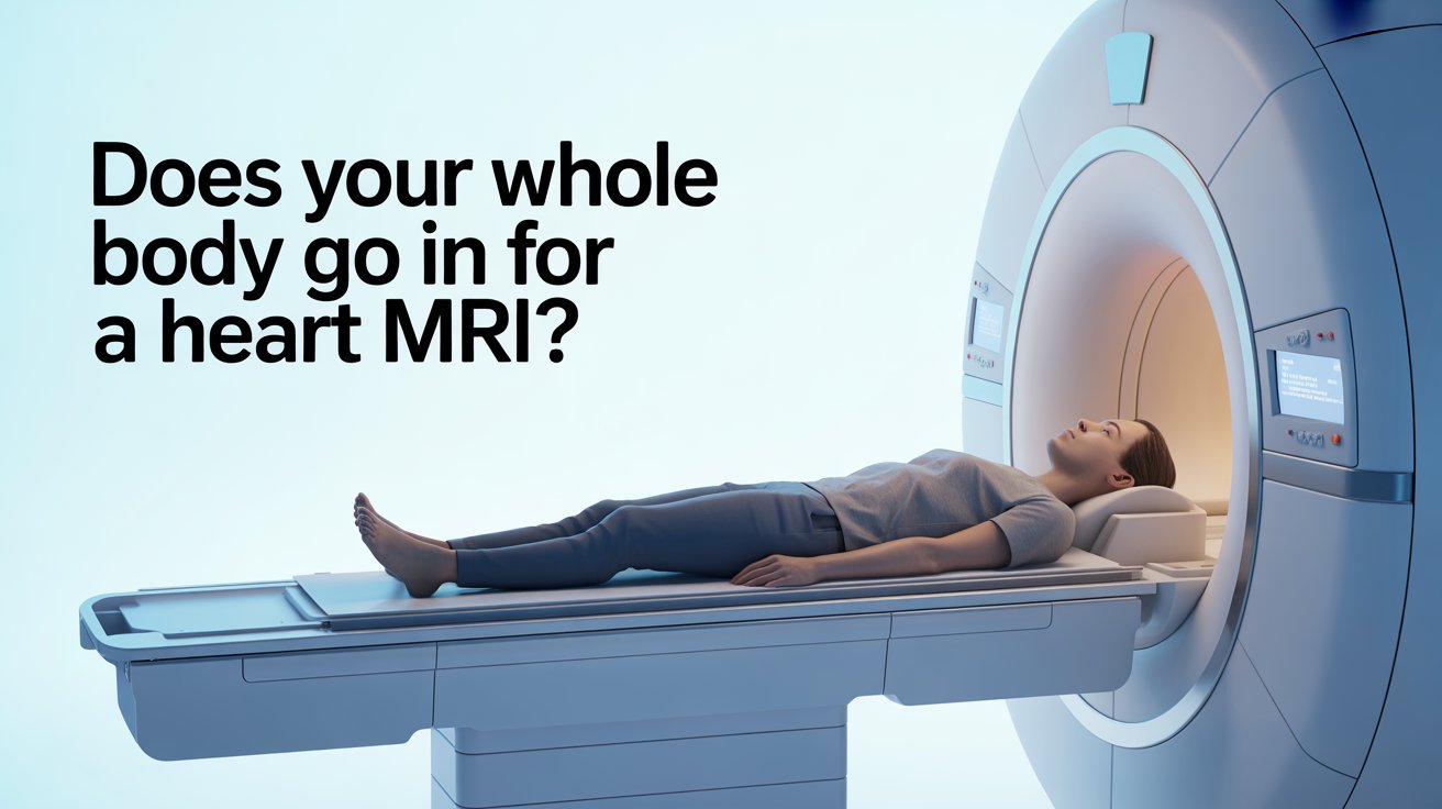
Does Your Whole Body Go Into the Scanner for a Heart MRI?

If you have been booked for a cardiac MRI and your biggest worry is whether you will be fully inside the scanner, the reassurance is this: you will not be. This is one of the most commonly searched questions before a cardiac MRI appointment — and the answer is simpler than most people expect.

A heart MRI focuses on the chest. That is the area the machine needs to image, and it is the area that enters the tunnel. Your head typically remains outside, and your legs and feet extend out the other end. The scanner is open at both ends throughout the entire scan.

If you are experiencing cardiac symptoms and want a prompt first assessment before being referred for advanced imaging, our ECG heart health check-up at The Private GP gives you same-day results with no waiting list.


Does Your Whole Body Go Into the Scanner?

No. For a cardiac MRI, only your chest and upper abdomen enter the tunnel. Your head stays outside at one end, and your legs and feet extend out the other.

Guy’s and St Thomas’ NHS Foundation Trust describes the MRI scanner as a large tube with a short, open tunnel through it. The tunnel is usually 60cm to 70cm wide and open at both ends. You lie on a bed that moves through the scanner — but for a cardiac scan, you slide in only far enough for your chest to be positioned within the imaging area.

Great Ormond Street Hospital NHS confirms that the MRI machine is shaped like a short, open-ended tunnel. The patient lies on a flat scanning bed that slides into the tunnel — but does not disappear entirely inside it.

For patients who are larger or broader across the shoulders, some centres offer a wider 80cm bore scanner. Guy’s and St Thomas’ advises patients who weigh more than 90kg to call ahead so the team can check which scanner is most appropriate for them.


What Happens During a Cardiac MRI?

You lie flat on a padded bed and are moved slowly into the scanner until your chest is positioned within the imaging area. The process is calm and methodical, and the radiographer communicates with you throughout via an intercom.

Guy’s and St Thomas’ NHS explains that ECG stickers are attached to your chest before the scan begins. These monitor your heartbeat in real time, allowing the machine to synchronise image capture with your heart rhythm — which is essential for producing clear, sharp cardiac images. If you have chest hair, a small area may need to be shaved to ensure the stickers adhere properly.

A blood pressure cuff may also be fitted to your arm and used to monitor your blood pressure at intervals during the scan.


What Does the Coil Do?

Before you enter the scanner, a flat pad called a coil is placed on your chest. Guy’s and St Thomas’ describes it as acting like an aerial for the scanner — it picks up the signals your body emits during imaging and converts them into higher quality pictures. A smaller pad called breathing bellows may also be placed on your abdomen to monitor your breathing pattern, which helps the machine capture certain images accurately.

You will be asked to hold your breath briefly at several points during the scan. The radiographer will tell you clearly when to hold and when to breathe normally. Most breath-holds last only a few seconds.

The scanner is noisy throughout — expect loud tapping and knocking sounds. Ear protection is always provided, and in most centres you can listen to music through headphones.


Will You Need Contrast Dye?

Not always. Contrast dye — a gadolinium-based agent injected through a small cannula in your arm — is used when the clinical team needs clearer images of blood flow, heart muscle scarring, or vessel detail. The British Heart Foundation explains that the dye makes images of blood flow to the heart show up more clearly on the scan.

If contrast is used, you will be advised to drink plenty of water afterwards to help flush the dye out of your system. If you have kidney problems, your doctor will check your kidney function before any contrast is given.


How Long Does a Heart MRI Take?

A standard cardiac MRI takes between 45 and 90 minutes, depending on the type of scan and the number of sequences needed.

University Hospital Southampton NHS confirms that you can expect your examination to take between 30 and 90 minutes depending on how complex your scan is. Guy’s and St Thomas’ advises allowing up to two hours for the full appointment, including preparation, the scan itself, and time afterwards if contrast or stress medication has been given.

A cardiac stress MRI — where medication is used to make your heart work harder, simulating the effect of exercise — takes longer than a standard scan, as two phases of imaging are required.

After the scan, a specialist analyses the images and prepares a written report, which is sent to the doctor who referred you.


What Conditions Can a Heart MRI Detect?

Cardiac MRI is one of the most comprehensive tests available for assessing the heart’s structure and function. It is often recommended when an echocardiogram (heart ultrasound) has not provided a complete enough picture.

The British Heart Foundation confirms that a cardiac MRI can help identify congenital heart disease, reduced blood flow to the heart muscle that may cause chest pain (angina), and other structural or functional abnormalities.

Conditions it can detect or help assess include:

Cardiomyopathy

Disease of the heart muscle — including hypertrophic, dilated, and arrhythmogenic types — where MRI shows the thickness, function, and scarring of the muscle in detail.

Heart attack scarring

Gadolinium contrast highlights areas of heart muscle damaged by a previous heart attack, distinguishing dead or scarred tissue from healthy muscle.

Congenital heart disease

Structural problems present from birth, such as holes between chambers or abnormal vessel connections, can be clearly mapped using cardiac MRI.

Heart valve disease

MRI can assess how well the valves are opening and closing and quantify the severity of any leakage or narrowing.

Pericardial disease

Inflammation or thickening of the sac surrounding the heart (the pericardium) shows up clearly on MRI.

Aortic abnormalities

Aneurysms, dissections, or narrowing of the aorta can be identified using MR angiography techniques within the same scan.


What Should You Know Before Your Cardiac MRI?

A few specific preparation steps apply to cardiac MRI that differ from other types of MRI.

Caffeine ban

Guy’s and St Thomas’ NHS advises avoiding all caffeine from midnight the night before your appointment. This includes coffee (regular and decaffeinated), tea, matcha, cola, and energy drinks. Caffeine raises the heart rate and interferes with image quality, particularly for stress perfusion scans.

Fasting

The American Heart Association advises that you may be asked not to eat or drink anything for four to six hours before the scan. Your appointment letter will confirm the exact requirement.

Metal and implants

Remove all jewellery, watches, and metal items before your appointment. If you have a pacemaker, implantable defibrillator, or any other metal device in your body, tell the cardiac MRI team as soon as possible. Many modern pacemakers are MRI-compatible, but this must be confirmed in advance.

Claustrophobia

If you are worried about the enclosed space, speak to your GP before the appointment. The American Heart Association confirms that a sedative can be prescribed to help you stay calm during the scan — but this must be arranged ahead of time. Some centres also offer wider bore scanners.

What to wear

Loose, comfortable clothing without metal zips or fasteners is ideal. A t-shirt and tracksuit bottoms or loose trousers are a practical choice.

If you are unsure whether a cardiac MRI is the right investigation for your symptoms, or if you need a referral, our private GP consultation can help you get the right answers quickly.


Frequently Asked Questions

  • Will my head go inside the MRI machine for a heart scan?

For most cardiac MRIs, no. Your chest enters the tunnel but your head typically remains outside. The scanner is open at both ends throughout the scan.

  • Is a cardiac MRI claustrophobic?

Some people find it uncomfortable, but most manage well. The scanner is open at both ends and your head stays outside. If you are anxious, a mild sedative can be prescribed by your GP before the appointment.

  • Can I have a cardiac MRI if I have a pacemaker?

Many modern pacemakers are MRI-compatible, but this must be confirmed before your appointment. Always inform the cardiac MRI team about any implant as early as possible so they can check and plan accordingly.

  • How long after a cardiac MRI will I get results?

A specialist analyses the images and sends a report to your referring doctor, typically within a few working days. Your doctor will then contact you to discuss the findings.

  • Is a heart MRI better than an echocardiogram?

They serve different purposes. An echocardiogram is quicker and often the first test used. Cardiac MRI provides more detailed images of heart muscle, scarring, and complex structures — and is typically recommended when more information is needed beyond what the echocardiogram showed.

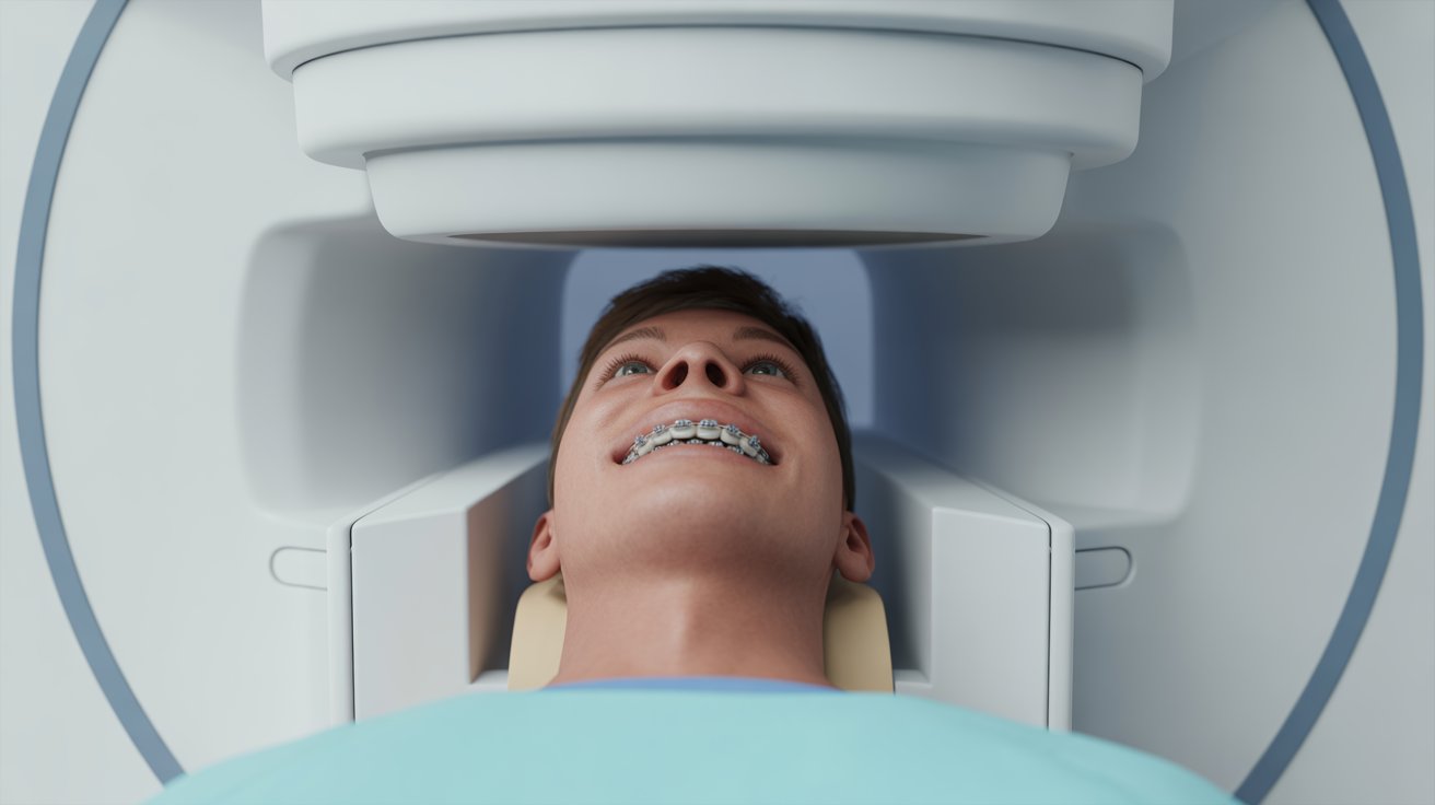
Can You Have an MRI With Braces?

Being referred for an MRI when you have braces is understandably confusing — the whole premise of the scan involves powerful magnets, and braces are made of metal. But the reassurance is straightforward: having braces does not prevent you from having an MRI.

Here is what you need to know before your appointment.


Is It Safe to Have an MRI With Braces?

Yes. Modern orthodontic braces are made from non-ferrous metal alloys — meaning they are not strongly attracted to magnets. They will not be pulled out of your mouth by the scanner, and they will not cause significant harm during the scan.

A clinical review published in the British Dental Journal confirms that orthodontic brackets are considered MRI-safe, though their proximity to the area being scanned can cause image artefact. The concern with braces is not patient safety — it is image quality near the mouth.

MRI Questions, a reference resource used by radiologists, confirms that metal orthodontic devices such as archwires and palate expanders can undergo some radiofrequency heating during a scan, but at levels far below the threshold that would cause pain or injury to the mouth or surrounding tissue.

That said, Princess Alexandra Hospital NHS Trust advises patients with fixed braces to contact the MRI department before their appointment. This is standard practice — not because the scan cannot go ahead, but so the team can plan appropriately and flag the potential for image distortion near the mouth.


Will Braces Affect the MRI Images?

Possibly — but it depends on which part of your body is being scanned.

The British Dental Journal review notes that metal orthodontic appliances are known to cause magnetic field distortions and signal loss, resulting in artefact on the images. In plain terms, the area close to the braces may appear blurred or distorted on the scan.

Zainab Clinics’ dental MRI safety guide explains that for scans of the brain, jaw, or sinuses — areas close to the mouth — the chances of image distortion are high and the radiologist needs to be aware. For scans of the abdomen, spine, knee, or lower limbs, braces are unlikely to affect the results at all.

Your radiologist will account for any artefact when reporting the images. In cases where a brain or jaw MRI is significantly affected, the team may discuss whether a repeat scan or alternative imaging is needed.


Does the Type of Brace Matter?

Yes. Different brace types have different implications for MRI.

Metal braces are the most common type and the most likely to cause image artefact near the mouth. They are still considered safe for scanning, but the MRI team should always be informed.

Ceramic braces are made from tooth-coloured ceramic material, which is non-metallic. They cause little to no interference with MRI images and are generally easier to scan with.

Clear aligners (such as Invisalign) are made entirely from plastic. They pose no problem for MRI and should be removed before entering the scanning room, as they sit loosely over the teeth.

Lingual braces — fitted to the back of the teeth — are made from similar metal alloys to standard braces and carry the same considerations. Declare them to the MRI team in the same way.


Do You Need to Tell the MRI Team About Your Braces?

Yes, always — regardless of which part of your body is being scanned.

Royal Devon University Healthcare NHS Foundation Trust’s MRI preparation guidance lists having a dental brace fitted as one of the conditions requiring patients to call the MRI department before attending. Princess Alexandra Hospital NHS Trust echoes this, asking patients with fixed braces to contact the department in advance.

This does not mean the scan will be refused or delayed. It simply allows the radiographer and reporting radiologist to prepare — noting that some artefact may appear near the mouth and adjusting their interpretation accordingly. Telling them in advance avoids any last-minute uncertainty on the day.

When you call or arrive, let the team know:

  • That you have fixed braces (or a fixed retainer)
  • Which type — metal, ceramic, or lingual
  • Which area of your body is being scanned

From there, the team will confirm whether any additional steps are needed.


What About Retainers and Clear Aligners?

Removable retainers and clear aligners should be taken out before entering the MRI scanning room. They are not a safety risk, but removing them is standard practice for any loose items in or around the mouth before a scan.

Fixed retainers — the thin wire bonded to the back of the front teeth after orthodontic treatment — are treated in the same way as fixed braces. They are considered MRI-safe, but you should declare them to the MRI team before your appointment. For most body scans, a fixed retainer causes no practical problem whatsoever.


Frequently Asked Questions

  • Will my braces be pulled out during an MRI?

No. Modern orthodontic braces are made from non-ferrous alloys that are not strongly attracted to the MRI magnet. There is no risk of them being pulled out during the scan.

  • Do I need to remove my braces before an MRI?

Fixed braces cannot be removed and do not need to be. Removable aligners and retainers should be taken out before entering the scanning room, as with any loose item near the mouth.

  • Can I have a brain MRI with braces?

Yes, though metal braces can cause some image distortion near the mouth. The radiologist will be aware of this and will account for it when interpreting the images. Tell the MRI department in advance so they can plan accordingly.

  • What should I tell the MRI team if I have braces?

Let them know you have fixed braces, what type they are, and which part of your body is being scanned. This allows the team to prepare and flag any potential artefact in their report.

  • Can I have an MRI with a fixed retainer?

Yes. A fixed retainer is treated the same way as fixed braces — considered MRI-safe, but worth declaring to the team before your appointment. For most body scans it causes no practical issue at all.

Can You Drive After an MRI Scan?

Whether you need to arrange a lift home from your MRI depends on one thing: whether you have been given a sedative. For the vast majority of patients having a standard scan, driving home afterwards is completely fine.

Here is the full picture, including what changes if you have had contrast dye or sedation.


Can You Drive After a Standard MRI?

Yes. A standard MRI scan has no known side effects and does not affect your ability to drive. You can leave the clinic and drive home immediately after the scan is complete.

Gateshead Health NHS confirms that for most MRI scans, there is no reason why you cannot drive home afterwards and resume normal activities. The MRI machine uses magnetic fields and radio waves — neither of which leave any residual effect in your body once the scan is finished.

You will be fully awake throughout the procedure, and the scan itself causes no drowsiness, impairment, or physical after-effects. Once the radiographer confirms you are free to leave, you can get in your car and go.


Can You Drive After an MRI With Sedation?

No. If you have been given a sedative before or during your MRI, you must not drive for 24 hours afterwards.

NHS Inform is clear that if you decide to have a sedative during the scan, you will need to arrange for a friend or family member to drive you home afterwards, as you will not be able to drive for 24 hours. It is also not safe to operate heavy machinery or drink alcohol during that 24-hour window.

Cambridge University Hospitals confirms the same guidance — if you have had a sedative, a friend or family member must take you home. You should not be left alone for the first 24 hours after sedation.

Sedation for an MRI is not given on the day — it must be prescribed by your GP well in advance of the appointment. If you think you may need one due to claustrophobia or anxiety, arrange this with your GP before your scan date so you have time to plan your transport home.


Can You Drive After an MRI With Contrast Dye?

In most cases, yes. Contrast dye alone does not affect your ability to drive.

Rotherham NHS Foundation Trust’s post-MRI patient guidance confirms that having MRI contrast media does not have any known effect on your ability to drive vehicles or operate machinery.

However, there is one important exception. York Hospitals NHS advises that if you experience any visual disturbance after your scan, you must not drive or operate machinery until your vision has fully returned to normal. Visual disturbance is a rare side effect of contrast dye, but if it occurs, it is a clear signal to stay put and inform the MRI team before you leave the department.

Most clinics will ask you to wait 15 minutes after a contrast injection before leaving, to monitor for any immediate reaction. Common mild side effects — such as a brief headache, slight nausea, or a warm sensation — typically pass within minutes. If you feel unwell after contrast, tell the MRI team before attempting to drive.


What If You Feel Unwell After Your MRI?

For the vast majority of patients, a standard MRI causes no after-effects whatsoever. If you do feel unwell after the scan, tell the radiology team before you leave — do not attempt to drive if you feel dizzy, nauseous, or unwell in any way.

If you received contrast dye and develop symptoms in the hours after leaving — such as a skin rash, wheezing, difficulty breathing, or a significant change in the amount of urine you are passing — seek medical advice promptly. Contact your GP or call NHS 111. In the event of a severe reaction, call 999.

Delayed allergic reactions to contrast dye are rare, but knowing the signs means you can act quickly if needed.


Frequently Asked Questions

  • Do I need to arrange a lift home from my MRI?

Only if you have been given a sedative. For a standard MRI — with or without contrast dye — you can drive yourself home immediately after the scan.

  • Can I drive after an MRI if I had contrast dye?

Yes, in most cases. Contrast dye has no known effect on driving ability. The exception is if you experience visual disturbances after the injection — in that case, do not drive until your vision has fully cleared.

  • How long after an MRI with sedation can I drive?

You must wait 24 hours after receiving a sedative before driving. Arrange a friend or family member to take you home, and do not be left alone for the first 24 hours.

  • Can I go back to work after an MRI?

Yes, for a standard MRI. You can return to work, drive, and carry on with your normal day immediately. If you had sedation, you should rest at home for the remainder of the day and not work for 24 hours.

  • Can I drink alcohol after an MRI?

After a standard MRI, yes. After a sedative, no — alcohol must be avoided for 24 hours following sedation, alongside driving and operating machinery.

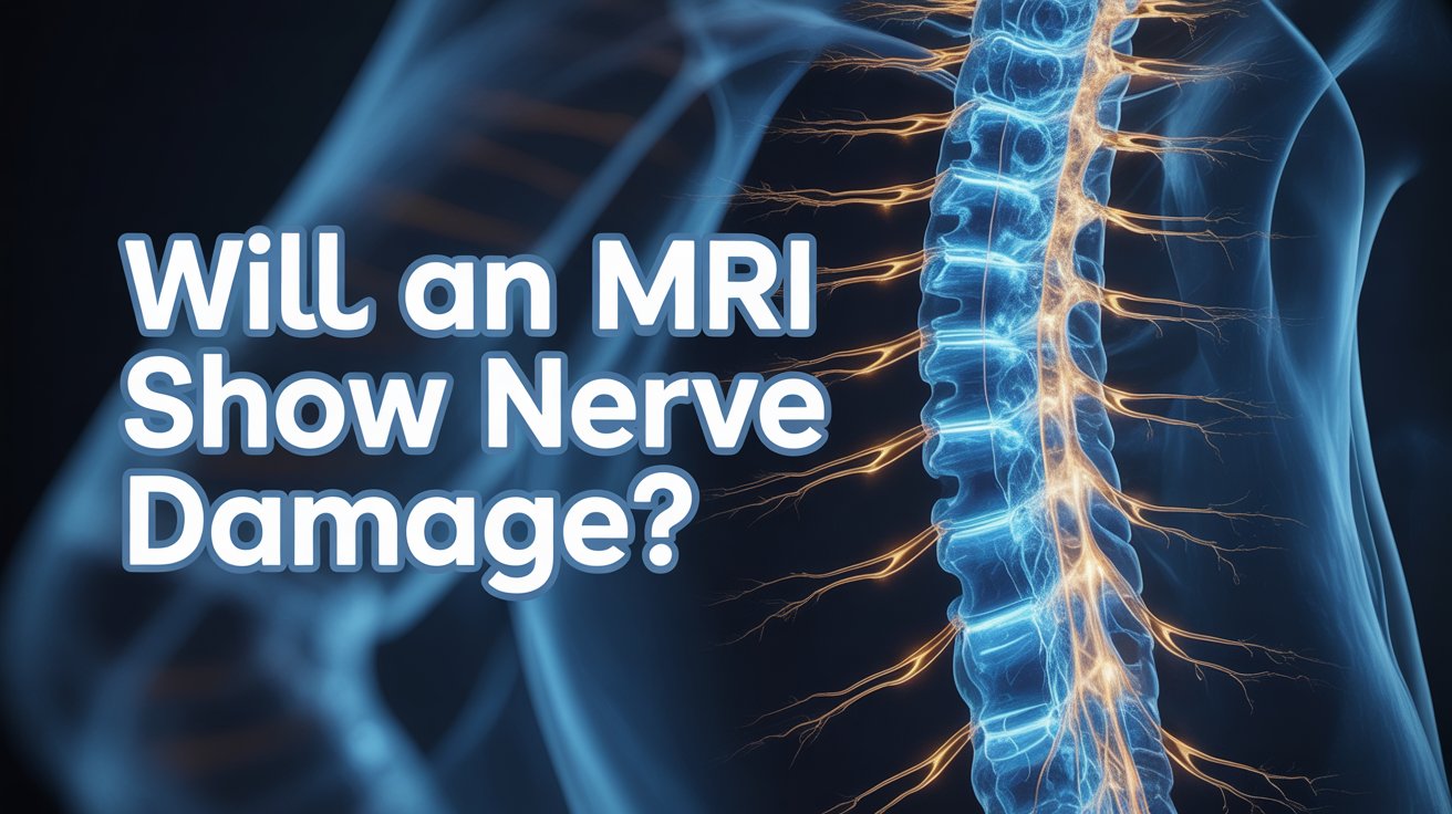
Will an MRI Show Nerve Damage?

Numbness, tingling, weakness, or burning pain are symptoms that drive thousands of people to ask whether an MRI will find the answer. The short answer is: often yes — but it depends on what type of nerve damage is involved and where it is in the body.

This guide explains what an MRI can and cannot show for nerve damage, when other tests are needed, and what symptoms should prompt you to seek assessment promptly.


Can an MRI Show Nerve Damage?

Yes. An MRI can detect nerve damage in many of the most common clinical situations — particularly where a nerve is being compressed, inflamed, or where surrounding structures are causing the problem.

Scan.com UK explains that MRI’s multiple views from different angles allow the easy detection of any displacement or protrusions putting pressure on a nerve, or anomalies in the area where the nerve exits the spine. It can also visualise the nerves themselves, along with conditions affecting them such as swelling or shrinkage.

What MRI does particularly well is show the structures surrounding nerves — the discs, vertebrae, ligaments, and soft tissues — in fine detail. When any of these press on, pinch, or damage a nerve, the MRI reveals it clearly. It also shows the spinal cord and nerve roots with a level of detail no X-ray can match.

Where MRI has limits is in measuring how well a nerve is actually working. It shows anatomy — what the nerve looks like and what is pressing on it — but not function. For that reason, it is sometimes used alongside other tests to build a complete picture.


What Types of Nerve Damage Does MRI Show Best?

MRI is most informative for nerve damage that has a clear structural or inflammatory cause. Here are the situations where it performs best.

Compressed spinal nerve roots

This is one of the most common reasons MRI is arranged for nerve symptoms. When a herniated disc, bone spur, or thickened ligament presses on a nerve root as it exits the spine, the MRI shows exactly which level is affected and how severely. This is the underlying cause of sciatica, cervical radiculopathy, and many cases of arm or leg pain, weakness, and numbness.

Spinal stenosis

Narrowing of the spinal canal compresses the spinal cord or multiple nerve roots simultaneously. MRI shows the degree of narrowing and which nerves are affected, informing decisions about treatment and surgery.

Multiple sclerosis (MS)

MS causes demyelination — damage to the protective sheath around nerve fibres. These lesions appear as bright white spots on T2-weighted MRI sequences, both in the brain and spinal cord. MRI is the primary tool for diagnosing and monitoring MS.

Spinal cord damage

Injury, inflammation, or reduced blood supply to the spinal cord itself — called myelopathy — produces characteristic changes on MRI that guide treatment urgently.

Brain-related nerve damage

Stroke, traumatic brain injury, and brain tumours can all disrupt nerve pathways. MRI of the brain identifies these structural causes with precision.


What Is MR Neurography?

For peripheral nerve damage — affecting the nerves beyond the spine — a specialist technique called MR neurography (MRN) provides significantly more detail than a standard MRI.

Research published in PMC confirms that MRN can detect small patches of inflammation in peripheral nerves and is valuable when standard MRI and nerve conduction studies have produced inconclusive results. It is particularly useful for assessing nerve lesions after trauma, planning surgery, and diagnosing entrapment neuropathies — such as carpal tunnel syndrome, ulnar nerve entrapment at the elbow, and sciatic nerve compression.

MRN uses high-resolution T2-weighted sequences with fat suppression to isolate the nerve signal from surrounding tissue, making individual peripheral nerves visible in a way that standard MRI cannot achieve. It is not available at every NHS centre and is more commonly accessed privately or through specialist neurological referral.


What Can MRI Not Show for Nerve Damage?

MRI shows structure, not function. This is its most important limitation when investigating nerve damage.

Howe Law’s clinical summary on MRI and nerve damage explains that MRI primarily shows the morphology of nerves — what they look like physically — but cannot provide definitive information about how well the nerve is working. A nerve can appear structurally normal on MRI while still conducting signals poorly or causing significant symptoms.

Specific types of nerve damage that standard MRI often cannot detect include:

Diabetic peripheral neuropathy

This condition damages small nerve fibres throughout the body, causing pain, numbness, and tingling — but often without the structural changes that MRI is designed to pick up. Howe Law notes that neuropathy secondary to diabetes may not always be detectable through standard MRI techniques.

Small fibre neuropathy

This affects the smallest sensory nerve fibres and typically requires a skin biopsy for diagnosis — MRI does not reliably show these microscopic changes.

Early-stage nerve damage

In the very early stages of compression or injury, structural changes may be too subtle to appear on MRI even while symptoms are already present.

A normal MRI result does not rule out nerve damage. If your symptoms are consistent with nerve involvement but your MRI is unremarkable, further investigation using functional tests is the appropriate next step.


What Other Tests Are Used to Diagnose Nerve Damage?

When MRI alone is not sufficient — or when the clinical question is about how well a nerve is working rather than what is pressing on it — two additional tests are commonly used.

Nerve Conduction Study (NCS)

A nerve conduction study measures the speed and strength of electrical signals travelling through peripheral nerves. North Bristol NHS Trust explains that it assesses the function of the sensory and motor nerve pathways, and is used when a patient is experiencing numbness, weakness, or changes in sensation to the hands or feet.

During the test, small electrical pulses are applied to the surface of the skin over a nerve. The response is recorded at another point along the nerve’s path. The speed and strength of that response reveal whether the nerve is conducting signals normally or is damaged.

Electromyography (EMG)

EMG measures the electrical activity inside muscles, revealing whether abnormal patterns are the result of nerve damage or a muscle disorder. King’s College Hospital NHS confirms that NCS and EMG together are useful for diagnosing entrapment syndromes such as carpal tunnel syndrome, polyneuropathy (for example in diabetic patients), and other peripheral nervous system disorders.

A fine needle electrode is inserted into the muscle being examined. The electrical activity it records — both at rest and during contraction — tells the clinician whether the nerve supplying that muscle is functioning normally.

NCS and EMG complement MRI rather than replace it. MRI shows the structural cause; NCS and EMG show the functional impact. Used together, they give clinicians the clearest possible picture of what is happening and why.


When Should You Get an MRI for Nerve Damage Symptoms?

If you are experiencing numbness, tingling, weakness, or pain that radiates from your neck or back into an arm or leg, an MRI is usually the right first-line imaging investigation. These symptoms suggest a nerve root is being compressed somewhere along the spine, and MRI is the most effective way to find out where and why.

Symptoms that warrant prompt assessment include pain, numbness, or tingling that travels down one arm or leg, unexplained muscle weakness, a sensation of burning or electric shocks in the limbs, and loss of coordination or balance.

Certain symptoms should be treated as urgent. Sudden or rapidly worsening weakness in both legs, loss of bladder or bowel control, or numbness in the groin and inner thighs (saddle anaesthesia) are red flag signs that require immediate emergency attention — call 999 or go to A&E.

For symptoms that are less acute — persistent but not rapidly worsening — a same-day private GP assessment allows for prompt clinical evaluation, appropriate investigation, and referral if needed. Our private GP consultation is available with no waiting list, and our full health check-up can help identify contributing factors such as diabetes or nutritional deficiencies that may be causing or worsening nerve symptoms.


Frequently Asked Questions

  • Can an MRI detect a pinched nerve?

Yes. MRI is one of the best tests for identifying a pinched nerve — it clearly shows if a disc, bone spur, or other structure is compressing a nerve root, and at which level of the spine.

  • Will an MRI show sciatica?

MRI can show the cause of sciatica — most commonly a herniated disc pressing on the sciatic nerve root. It does not diagnose sciatica itself, but reveals the structural problem driving the symptoms.

  • Can an MRI show nerve damage in the neck?

Yes. A cervical spine MRI shows the nerve roots exiting the neck, any disc herniation or stenosis compressing them, and the spinal cord itself. It is the standard investigation for neck-related arm pain, numbness, and weakness.

  • What is the best test to diagnose nerve damage?

It depends on the type. MRI is best for structural causes such as disc compression and MS. Nerve conduction studies and EMG are best for assessing peripheral nerve function. In many cases, a combination of tests gives the clearest diagnosis.

  • Can an MRI miss nerve damage?

Yes. MRI shows structure, not function, so it can appear normal even when nerve damage is present — particularly in diabetic neuropathy, small fibre neuropathy, and early-stage compression. A normal MRI does not rule out nerve damage if symptoms persist.

How Long Does a Full Spine MRI Take?

A full spine MRI covers a lot of ground — from the base of your skull all the way down to your lower back. That means it takes considerably longer than a scan of just one section. If you know what to expect time-wise before you arrive, the appointment is far less daunting.

This guide covers how long the scan takes, how it compares to individual section scans, what happens during the appointment, and what a full spine MRI can show.


How Long Does a Full Spine MRI Take?

A full spine MRI typically takes between 60 and 90 minutes inside the scanner. You should allow up to two hours for your total time at the clinic, including preparation, changing, safety checks, and any post-scan wait if contrast dye is used.

Healthline confirms that a cervical spine scan takes around 20 to 45 minutes, and a lumbar spine scan takes around 30 to 60 minutes. A full spine scan — covering all three sections — requires each region to be imaged separately with its own sequences, which is why the total time is significantly longer than any individual section alone.

Harrogate and District NHS Foundation Trust notes that while most MRI scans take around 30 minutes, some take up to 90 minutes — and a full spine scan sits firmly at the longer end of that range.


How Does This Compare to a Single Section Scan?

Here is a quick breakdown of typical scan times by spinal region:

  • Cervical spine (neck): 20 to 45 minutes
  • Thoracic spine (mid-back): 30 to 45 minutes
  • Lumbar spine (lower back): 30 to 60 minutes
  • Full spine (all three sections): 60 to 90 minutes

Each section requires the coil to be repositioned and a new set of sequences to be run, which is why three sections together do not simply equal three times as long — but they do add up considerably.


What Happens During a Full Spine MRI?

You lie flat on a padded bed and enter the scanner head-first. University Hospital Southampton NHS confirms that you will be positioned in the centre of the scanning bed and will need to lie very still throughout to achieve the best quality imaging.

A coil — a flat receiver pad — is placed over the section of your spine being imaged. For a full spine scan, the radiographer will reposition this coil between sections, which adds a small amount of time between imaging runs. You will be moved out briefly, the coil adjusted, and then moved back in for the next section.

The scanner produces loud tapping and knocking sounds throughout. University Hospital Southampton advises that you may also feel your body temperature increase slightly and experience tingling in your hands and feet — both of which are normal sensations caused by the magnetic field. You will be given an emergency buzzer to hold at all times, and the radiographer can hear you via a built-in microphone system between sequences.

For a lumbar spine scan specifically, Ezra’s clinical guide notes that patients are often positioned feet-first rather than head-first, which can make the experience more comfortable for those who feel anxious about enclosed spaces — as the head and upper body remain outside the tunnel for that section.


What Can Make a Full Spine MRI Take Longer?

Several factors can extend the time beyond the typical 60 to 90 minutes.

Contrast dye

If your clinician has requested contrast-enhanced imaging, a gadolinium-based dye is injected through a cannula in your arm. A second set of images is taken after injection, adding around 15 to 30 minutes to the scan time.

Movement

The MRI machine is sensitive to any movement. UK Spine Centre confirms that it is crucial for patients to remain still throughout the scanning process to ensure clear images. If movement blurs a sequence, it must be repeated — which extends the total time.

Number of sequences

The number of image sequences ordered by your clinician varies depending on what they are investigating. More detailed or complex clinical questions require more sequences, which adds time.

Patient size

Larger body habitus can require adjustments to coil positioning and signal strength settings, which occasionally adds time to individual sequences.


What Does a Full Spine MRI Show?

A full spine MRI gives clinicians a comprehensive view of the entire spinal column — from the vertebrae and discs to the spinal cord, nerve roots, and surrounding soft tissue.

UK Spine Centre outlines the range of conditions a spinal MRI can identify, including:

Disc herniation and sciatica

A herniated disc pressing on a nerve root is one of the most common reasons for a spinal MRI. The scan shows exactly which disc is affected and how severely it is compressing the nerve.

Spinal stenosis

Narrowing of the spinal canal — whether from disc degeneration, bone spurs, or thickened ligaments — compresses the spinal cord or nerve roots and shows up clearly on MRI.

Nerve compression

Wherever a nerve is being pinched or squeezed along the length of the spine, MRI identifies the cause and the level of involvement.

Spinal cord abnormalities

Conditions affecting the cord itself — including MS lesions, inflammation, or areas of poor blood supply — are visible on MRI in a way no other imaging method can match.

Tumours

Both primary spinal tumours and metastatic disease (cancer that has spread to the spine) can be identified and characterised using MRI.

Fractures and infections

Vertebral fractures — including stress or compression fractures that may not appear on X-ray — and bone infections (osteomyelitis) show up on MRI with detail that guides treatment decisions.

Post-surgical assessment

If you have had previous spinal surgery, MRI can assess whether there is scarring, recurrent disc herniation, or infection around the operated area.


How to Prepare for a Full Spine MRI

Preparation for a full spine MRI is straightforward. Ezra’s guidance confirms that unless told otherwise, you can eat, drink, and take your usual medications as normal on the day. No fasting is required for a standard spine MRI without contrast. If contrast dye has been requested, your appointment letter will specify any fasting instructions.

Remove all jewellery, piercings, and metal accessories before attending. Wear loose, comfortable clothing without metal zips, buttons, or fasteners — a t-shirt and tracksuit bottoms work well. You may be asked to change into a hospital gown on arrival.

Tell the MRI team in advance about any metal implants, surgical clips, or devices in your body, including spinal hardware such as rods, screws, or cages from previous surgery. Most modern spinal implants are MRI-compatible, but this must be confirmed beforehand.

If you are anxious about lying still for 60 to 90 minutes or feel uncomfortable in enclosed spaces, speak to your GP before the appointment. A mild sedative can be prescribed to help you remain calm, but it must be arranged in advance as it cannot be prescribed on the day.

If you are experiencing back, neck, or leg symptoms and are unsure whether a spine MRI is the right investigation, our full health check-up gives you a thorough clinical assessment to help determine the most appropriate next step.


Frequently Asked Questions

  • Is a full spine MRI the same as three separate scans?

Effectively yes — each section (cervical, thoracic, lumbar) is imaged with its own sequences. It is done in one appointment, but the coil is repositioned between sections and each region is scanned individually.

  • Can I move between sections of a full spine MRI?

You will be briefly moved out of the scanner between sections for coil repositioning. During each imaging run, you must remain completely still to avoid blurred images requiring repetition.

  • Will my whole body go inside the scanner for a full spine MRI?

For the cervical and thoracic sections, most of your body enters the tunnel. For the lumbar section, entry is often feet-first, meaning your head and upper body can remain outside.

  • How long after a full spine MRI will I get results?

A radiologist analyses the images and sends a report to your referring doctor, typically within a few working days. Your doctor will then contact you to discuss the findings.

  • Do I need contrast dye for a full spine MRI?

Not always. Contrast is used when the clinical team needs clearer detail — for example, when investigating tumours, infection, or inflammation. Your appointment letter will confirm whether it has been requested.

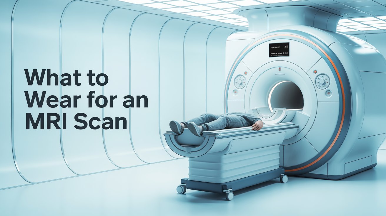
What to Wear for an MRI Scan

Choosing the right outfit for an MRI takes about two minutes of planning and can make your appointment considerably more comfortable. The main rule is simple: avoid metal. Here is everything you need to know before you arrive.


What Should You Wear to an MRI Scan?

Wear loose, comfortable clothing with no metal fastenings. This is the single most important clothing rule for an MRI, and it applies regardless of which part of your body is being scanned.

NHS Inform confirms that if you do not need to wear a gown, you should wear clothes without metal zips, fasteners, buttons, underwire bras, belts, or buckles. Surrey and Sussex Healthcare NHS Trust advises choosing thin cotton-based clothing with minimal layers, as this also helps with cooling during the scan — the magnetic field can cause a mild warming sensation in some patients.

Good choices include:

  • A plain t-shirt or cotton top with no metal press studs or zips
  • Loose tracksuit bottoms or joggers with an elasticated waistband
  • Soft, wireless sports bra or a comfortable bralette
  • Slip-on shoes or trainers without metal eyelets


What Clothing to Avoid

Certain everyday items cause problems in the scanning room and should be left at home or be prepared to be removed:

Jeans

Most jeans have metal rivets, metal zips, and metal buttons. They are one of the most common items patients are asked to remove before a scan.

Underwired bras

The metal underwire must be removed before scanning. A wireless bra or soft sports bra is a much easier choice and saves the need to change.

Belts

Any belt with a metal buckle needs to come off. Elasticated waistbands avoid this entirely.

Hoodies and jackets

Many have metal zips, metal drawstring tips, or metal poppers. A plain sweatshirt or cardigan without fastenings is a better option.

Compression garments

Some compression leggings and athletic wear contain metallic fibres in the fabric. If in doubt, opt for plain cotton instead.

Leeds Teaching Hospitals NHS Trust advises that if there is any metal on your clothing near the area being scanned, you may be asked to remove that item of clothing and a gown will be provided.


Does It Matter Which Body Part Is Being Scanned?

Yes, slightly. The area being scanned determines how much of your clothing is relevant.

For head, brain, or neck scans, your clothing from the shoulders down is generally not a concern. Focus on removing all metal from the head and neck area — hairpins, earrings, necklaces, and piercings.

For chest or cardiac scans, avoid underwired bras and anything with metal across the chest. ECG stickers are attached to the chest during cardiac MRI, so easy access matters. If you are having an ECG heart health check-up as part of your cardiac assessment, the same principle applies.

For spine, abdomen, or pelvic scans, the focus shifts to your lower half. Avoid jeans, belts, and anything with metal fastenings around the waist or hips. Loose trousers or joggers are the simplest choice.

For knee, ankle, or foot scans, most of your clothing is irrelevant to the scan itself. Wear whatever is comfortable and easy to remove from the lower leg if needed.


Will You Need to Change Into a Hospital Gown?

Sometimes — but not always. Whether you need a gown depends on which part of your body is being scanned and whether your clothing is already metal-free.

NHS Inform confirms that depending on which part of your body is being scanned, you may need to wear a hospital gown during the procedure. The Royal Marsden NHS Trust advises that when you arrive for your appointment, it is advisable to wear clothing without metal fastenings — alternatively, they can provide a hospital gown or pyjamas.

If you arrive in entirely metal-free clothing, many clinics will allow you to stay in your own clothes throughout. This is one of the reasons arriving dressed appropriately saves time and avoids any awkwardness on the day.

When a gown is provided, you will be directed to a changing room with a secure locker for your belongings. Your dignity and privacy are maintained throughout — only the area being scanned is exposed, and you remain covered otherwise.


What Jewellery and Accessories Should You Remove?

All of it. Every piece of metal jewellery and every metal accessory must be removed before you enter the scanning room, without exception.

This includes:

  • Rings, earrings, necklaces, and bracelets
  • Body piercings of any kind
  • Watches and fitness trackers
  • Hair clips, kirby grips, and metallic hairbands
  • Hearing aids
  • Glasses and spectacle frames
  • Removable dental work such as retainers or partial dentures

Royal Devon University Healthcare NHS Foundation Trust advises attending with clothing that has no clips, zips, or belt fastenings, and removing all metal accessories before the scan.

Leave valuables at home where possible. Most clinics provide secure lockers for items you do bring, but a lost or damaged item of jewellery or a watch with a damaged magnetic strip is easily avoided by not bringing it in the first place. Credit and debit cards should also be left outside the scanning room, as the magnetic field can erase or damage the card’s strip.


What About Make-Up and Cosmetics?

Make-up is a less obvious consideration, but it is worth knowing about before your appointment.

Leeds Teaching Hospitals NHS Trust specifically advises not wearing cosmetics with magnetic substances — including magnet-attached eyelashes — and ensuring any previously applied make-up of this type is fully removed on the day. Royal Devon NHS advises avoiding heavy eye make-up, as this can affect the quality of some scans.

Some mascaras, eyeshadows, and eyeliners contain iron oxide or other metallic pigments. These particles can interact with the magnetic field, causing a mild warming sensation around the eye area and occasionally affecting image quality near the head.

Magnetic eyelashes — which use tiny magnets as their adhesive mechanism — must be removed before scanning without exception. Standard press-on lashes attached with glue are fine.

Skin lotions, moisturisers, and sun cream do not pose a problem for most MRI scans. However, for cardiac MRI where ECG electrodes are attached to the chest, any lotion applied to the chest area should be washed off beforehand, as it can reduce electrode adhesion and affect monitoring quality.


Frequently Asked Questions

  • Can I wear a sports bra for an MRI?

Yes, provided it is wireless and has no metal clasps, hooks, or underwire. A soft, wireless sports bra or bralette is the ideal choice for any scan involving the chest or upper body.

  • Can I wear earrings to an MRI?

No. All earrings — including small studs — must be removed before entering the scanning room. If you have a new piercing that cannot be removed, let the MRI team know in advance so they can advise.

  • What happens if I forget and wear metal to my MRI?

You will be asked to remove the item before the scan. If it is an item of clothing that cannot easily be removed, a gown will be provided. The scan will still go ahead in most cases — it just adds a few minutes to your preparation time.

  • Can I wear my own clothes or do I have to wear a gown?

If your clothing is entirely metal-free, many clinics will allow you to stay in your own clothes. If there is any metal near the area being scanned, a gown will be provided. Arriving in suitable clothing is the easiest way to avoid needing to change.

  • Can I wear nail varnish to an MRI?

Standard nail varnish is fine for most MRI scans. Some metallic or glitter nail polishes contain metal particles and may cause a slight warming sensation, but this is uncommon. If you are having a scan of the hands or fingers, it is worth removing nail varnish beforehand to avoid any interference with image quality.

Can You Eat Before an MRI Scan?

You have an MRI booked and you are wondering whether you need to skip breakfast. For most people, the answer is no. But the rules vary depending on what is being scanned and whether contrast dye is involved — and getting this wrong could mean your scan has to be postponed and rescheduled.

This guide gives you a clear answer based on your scan type, explains why fasting is sometimes necessary, and covers everything else you need to know to walk into your appointment prepared.


Can You Eat Before an MRI? The General Rule

For most standard MRI scans, you can eat, drink, and take your usual medication as normal beforehand. No special preparation is needed.

The NHS confirms that the hospital where you are having your MRI will tell you if there is anything you need to do before the scan. If your appointment letter does not mention fasting or dietary restrictions, you do not need to change anything.

Cambridge University Hospitals’ patient information is explicit: on the day of your MRI scan, you should be able to eat, drink, and take any medication as usual, unless you have been advised otherwise. Leeds Teaching Hospitals confirms the same — in most cases you do not need to do anything to prepare for the scan and can eat and drink as normal.

This means that if you are having a brain MRI, a spinal MRI, a knee scan, a shoulder scan, or any other joint or extremity scan, you can have your breakfast, lunch, or dinner as normal before you arrive. There is no need to rush out of the house on an empty stomach.

The one golden rule that applies to every MRI appointment: read your appointment letter carefully and follow any specific instructions it contains. If the letter says to fast, fast. If it says nothing about food, eat as normal. If you are ever unsure, ring the radiology department before the day.


When Do You Need to Fast Before an MRI?

Fasting is required for a specific set of MRI scans — those where food in the digestive system would blur the images, or where contrast dye or sedation creates a nausea or safety risk.

Ezra’s clinical guide on MRI preparation explains that although many MRI scans can proceed without prior fasting, a sub-selection of scans require it. Here are the main situations where you will be asked to fast.

Abdominal and pelvic MRI

When the scanner is imaging your abdomen — including the liver, pancreas, kidneys, gallbladder, and surrounding organs — food in your digestive system causes a problem. Eating triggers peristalsis, the involuntary muscle contractions that move food through your gut. These movements create motion on the images, known as artefacts, which blur the scan and can obscure abnormalities. Fasting slows this activity down, giving the radiologist a much clearer picture. University Hospitals Plymouth’s liver MRI patient information confirms that patients must not eat anything for 4 hours before their appointment, though they can drink water or squash in that time.

Liver, pancreas, and gallbladder scans

As well as the motion artefact problem, eating causes your gallbladder to contract and shrink, making it harder for the radiologist to assess it properly. Fasting keeps the gallbladder distended and clearly visible.

MRCP (Magnetic Resonance Cholangiopancreatography)

This is a specialist MRI used to image the bile ducts, gallbladder, and pancreatic duct system. The fasting requirement for MRCP is particularly strict, often 6 to 8 hours, because the scan requires the digestive system to be as empty as possible to avoid fluid secretions interfering with the images.

MRI with contrast dye

When a gadolinium-based contrast agent is injected during the scan, it can occasionally cause nausea as a side effect. GetScanned’s fasting guide explains that fasting for 4 to 6 hours before a contrast scan is usually required to reduce the risk of vomiting while you are lying flat inside the scanner — a potentially serious situation as stomach contents could enter the airway. Your radiology department will specify the exact fasting period in your appointment instructions.

Cardiac MRI

For certain cardiac MRI examinations, food restriction and caffeine avoidance are both required. Craft Body Scan’s clinical preparation guide confirms that caffeine — including coffee (regular and decaf), tea, energy drinks, and chocolate — must be avoided for 24 hours before a cardiac MRI. Caffeine increases heart rate, making it harder to capture clear, stable images of the heart. If you are investigating cardiac symptoms, our ECG heart health check-up is a useful first step before moving to advanced cardiac imaging.

Scans requiring sedation or general anaesthetic

If you are having a sedative to manage claustrophobia or anxiety, or if your scan requires a general anaesthetic, fasting is essential for safety. When sedated, the protective reflexes that prevent aspiration — inhaling stomach contents if you vomit — are reduced. Fasting ensures the stomach is empty if this occurs. The Royal Marsden NHS Trust confirms that all instructions will be included in your appointment letter, and it is important to read it carefully.


How Long Do You Need to Fast Before an MRI?

When fasting is required, the standard period is 4 hours — no food, though water is usually still permitted.

NHS Highland’s patient information for fasting MRI scans gives a clear instruction: do not eat any food or drink any fluids for 4 hours before your appointment time. Cambridge University Hospitals confirms that in some cases you may be asked not to eat or drink anything for up to four hours before the scan.

For MRCP and some gastrointestinal MRI scans, the fasting period may be longer — up to 6 to 8 hours — so that the digestive system is as empty and still as possible. Your appointment letter will specify the exact time.

If you have been asked to fast and are finding it difficult, try to book an early morning appointment where possible. Sleeping through most of the fasting window makes it considerably more manageable. If you have diabetes or another condition that makes fasting medically complicated, contact the radiology department before your appointment — this is covered in more detail below.


Can You Drink Water Before an MRI?

In most cases, yes — water is permitted before an MRI, even during a fasting period.

GetScanned’s fasting preparation guide confirms that water is often allowed before an MRI and that staying hydrated is actually recommended, as it can make vein access easier if a contrast injection is needed. University Hospitals Plymouth’s liver MRI leaflet specifically tells patients they may drink water or squash in the 4 hours before their scan, even though food is not permitted.

Some pelvic MRI scans actually require you to arrive with a full bladder. In those cases, you will be asked to drink a specified amount of water before your appointment and not to urinate until after the scan is complete. Your appointment letter will tell you if this applies to you.

One important exception: caffeine. For cardiac MRI specifically, Craft Body Scan’s guide confirms that coffee (including decaffeinated), tea, energy drinks, cola, and chocolate must all be avoided for 24 hours before the scan. These contain caffeine which raises the heart rate and makes clear cardiac imaging significantly harder. If in doubt, stick to water for the day before a cardiac MRI.

A practical note worth keeping in mind: MRI scans can last between 30 minutes and 90 minutes, and you cannot leave the scanner partway through. It is sensible to limit how much you drink in the hour or two immediately before your appointment, simply for your own comfort.

After your scan, GetScanned advises drinking plenty of water — especially if you had contrast dye — to help your kidneys flush the gadolinium from your system efficiently.


Can You Take Medication Before an MRI?

For most patients, the answer is yes — continue taking all your regular medication as normal before an MRI.

Cambridge University Hospitals and Leeds Teaching Hospitals both confirm that in most cases you should eat, drink, and take your medication as usual. Royal United Hospital Bath’s MRI patient leaflet states that you should continue to take all your normal medication, with one important exception.

Diabetic patients need to take extra care, particularly those taking insulin or metformin. If you have diabetes and your MRI requires fasting, the interaction between fasting and your diabetes medication needs to be managed carefully. NHS Highland’s fasting MRI guidance includes specific diabetic instructions: if you take diabetes medication with food as prescribed, adjustments will be needed around the fasting window. Contact the radiology department before your appointment to discuss this — most NHS trusts aim to book diabetic patients for early-morning slots to minimise the fasting period.

If you have kidney disease and are taking metformin, this is particularly important to mention before any MRI involving contrast dye. GetScanned’s guide advises that metformin can pose a risk of complications in individuals with kidney problems when contrast is used. Our private blood tests can check your kidney function promptly if this is a concern before your scan.

Sedatives for claustrophobia must be prescribed by your GP before your appointment — the radiology department cannot prescribe medication on the day. If you think you might struggle with the enclosed space, speak to your GP well in advance. If you do take a sedative, you will need someone to drive you home afterwards, as you will not be able to drive for 24 hours.


Other Things to Know Before Your MRI Appointment

Beyond the question of food, there are a few other practical points worth knowing before your scan.

Metal

Remove all jewellery, piercings, watches, and accessories before you arrive. Avoid underwire bras, belts with metal buckles, and clothing with metal zips or buttons near the area being scanned. Leeds Teaching Hospitals suggests arriving in a t-shirt, jogging bottoms, and a sports bra if possible, to avoid having to change into a hospital gown.

Make-up

Some mascaras and metallic make-up products contain iron particles that can cause interference with image quality or feel warm during the scan. NHS Highland’s preparation guidance specifically advises patients not to wear mascara on the day. If in doubt, go make-up free for the appointment.

Glucose monitors

Leeds Teaching Hospitals’ patient information notes that most manufacturers advise removing continuous glucose monitors before an MRI. The device poses no safety risk to you, but the strong magnetic field may damage the sensor. Bring a replacement sensor patch with you so you can reapply it after the scan.

Tattoos

Some tattoos contain traces of metallic pigment. Let the radiographer know if you have tattoos near the area being scanned. Most are entirely unaffected, but very occasionally a tattoo may feel slightly warm during the scan.

Your appointment letter

This is the most important piece of preparation. Read it carefully before the day — it will tell you whether fasting is required, whether you need to drink water, whether you should avoid any specific medications, and what to bring. If your letter does not mention fasting, you do not need to fast. If anything is unclear, call the radiology department directly.

A full health check-up at The Private GP can help you understand your overall health picture, including whether an MRI is the right investigation for your symptoms and what other tests might be useful alongside it.


Frequently Asked Questions

  • Can I have coffee before an MRI?

For most standard MRI scans, yes — a cup of coffee before your appointment will not affect the scan. However, for cardiac MRI specifically, caffeine must be avoided for 24 hours before the examination. Craft Body Scan’s clinical guide confirms that coffee (including decaffeinated), tea, energy drinks, and chocolate all need to be avoided before cardiac imaging, as caffeine raises the heart rate and interferes with image quality. Check your appointment letter to confirm whether this applies to you.

  • What happens if I eat before an MRI when I was told to fast?

Tell the radiology team as soon as you arrive. Depending on what you ate, how long ago, and the type of scan, they may be able to go ahead — or they may need to reschedule. Eating before an abdominal or pelvic scan can result in blurred images that cannot be properly interpreted, meaning the scan would need to be repeated regardless. If you ate before a contrast scan, the team will assess the risk of nausea and advise accordingly. It is always better to tell them than to stay silent.

  • Can I eat before an MRI with contrast dye?

Generally no — or at least not in the 4 to 6 hours immediately before. GetScanned’s fasting guide confirms that fasting before a contrast MRI is usually required to reduce the risk of nausea and vomiting while you are lying flat inside the scanner. Your appointment letter will specify the exact fasting window. Water is usually permitted during the fasting period.

  • Can I eat normally before a brain MRI?

Yes. A brain MRI does not require any dietary preparation. Cambridge University Hospitals’ patient information confirms that you should eat, drink, and take your medication as usual on the day of a standard brain scan unless specifically advised otherwise. You can have a full meal beforehand and eat normally after the scan too.

  • What can I eat after an MRI scan?

There are no dietary restrictions after a standard MRI. The Royal Marsden NHS Trust confirms that you can eat and drink as usual as soon as your scan is finished. If you had contrast dye, drink plenty of water afterwards to help your kidneys flush the gadolinium from your system. If you received sedation, you may want to wait until you feel fully alert before eating a full meal.